
Depressive and Neurocognitive Disorders in the Context of the Inflammatory Background of COVID-19


Department of Psychiatry, Medical University of Bialystok, pl. Brodowicza 1, 16-070 Choroszcz, Poland
*
Author to whom correspondence should be addressed.
Life 2021, 11(10), 1056; https://doi.org/10.3390/life11101056
Submission received: 3 September 2021
/
Revised: 1 October 2021
/
Accepted: 3 October 2021
/
Published: 8 October 2021
(This article belongs to the Special Issue Depressive Disorders-New Challenges)
Abstract
:The dysfunctional effects of the coronavirus disease 2019 (COVID-19) infection on the nervous system are established. The manifestation of neuropsychiatric symptoms during and after infection is influenced by the neuroinvasive and neurotrophic properties of SARS-CoV-2 as well as strong inflammation characterised by a specific “cytokine storm”. Research suggests that a strong immune response to a SARS-CoV-2 infection and psychological stressors related to the pandemic may cause chronic inflammatory processes in the body with elevated levels of inflammatory markers contributing to the intensification of neurodegenerative processes. It is suggested that neuroinflammation and associated central nervous system changes may significantly contribute to the etiopathogenesis of depressive disorders. In addition, symptoms after a COVID-19 infection may persist for up to several weeks after an acute infection as a post-COVID-19 syndrome. Moreover, previous knowledge indicates that among SSRI (selective serotonin reuptake inhibitor) group antidepressants, fluoxetine is a promising drug against COVID-19. In conclusion, further research, observation and broadening of the knowledge of the pathomechanism of a SARS-CoV-2 infection and the impact on potential complications are necessary. It is essential to continue research in order to assess the long-term neuropsychiatric effects in COVID-19 patients and to find new therapeutic strategies.
1. Introduction
At the time of writing this review, the entire world was struggling with the coronavirus disease 2019 (COVID-19) pandemic, which has affected nearly every aspect of society [1]. Despite the duration of the pandemic for many months, SARS-CoV-2 infection still hides many unknowns, especially regarding its long-term effects [2,3]. COVID-19 is an acute infectious respiratory disease caused by a novel virus named SARS-CoV-2. It is a single-stranded RNA virus with about 80% similarity to SARS-CoV from the coronavirus family [4]. The first cases of infection were reported at the turn of 2019/2020 in China. Rapid global transmission of the virus was observed with an increasing wave of infections and deaths. In March 2020, The World Health Organization (WHO) declared a COVID-19 pandemic [5]. As of 30 August 2021, the COVID-19 pandemic had over 216 million confirmed cases with 4.5 million deaths [6]. Along with the destabilisation of everyday life, negative psychological consequences of the pandemic and the emergence of mental disorders among societies have been observed [7,8,9]. In the course of a COVID-19 infection, in addition to typical respiratory symptoms, a dysfunctional effect of SARS-CoV-2 on other body systems, including the nervous system, has been noticed [10,11]. A range of COVID-19 neurological symptoms has been observed from mild symptoms such as a headache or dizziness to loss of taste, smell, meningitis and even necrotic encephalitis [12,13]. According to the latest scientific reports, the manifestation of neuropsychiatric symptoms during and after a COVID-19 infection is influenced by the neuroinvasive and neurotrophic properties of SARS-CoV-2 as well as by strong inflammation in the course of the infection characterised by a specific “cytokine storm” [14,15,16]. Potentially life-threatening cytokine storm syndrome is characteristic of severe COVID-19 [17]. Research suggests that a strong immune response to a SARS-CoV-2 infection and psychological stressors related to the pandemic may cause chronic inflammatory processes in the body with elevated levels of inflammatory markers contributing to the intensification of neurodegenerative processes and the emergence of other psychiatric complications [18,19,20,21]. Inflammation, related changes and damages at the level of the nervous system in the course of SARS-CoV-2 have been suggested to significantly contribute to the etiopathogenesis of depressive disorders [22,23,24]. Furthermore, studies show that some acute viral infections can induce an abnormal immune response, affecting the manifestation of long-term neuropsychiatric consequences [25,26]. Experiences of previous viral pandemics, such as the flu of the 18th and 19th centuries, Spanish flu in the 20th century and the coronaviruses causing severe acute respiratory syndrome (SARS) in 2002 and the Middle East respiratory syndrome (MERS) in 2012, suggest a link with the manifestation of neuropsychiatric complications both during and after an acute infection [27]. Among the survivors after infection with SARS-CoV and MERS coronaviruses, there have been cases of memory, attention and concentration disturbances lasting up to 39 months after the onset of the disease [28,29]. It has been suggested that the severity of neuropsychiatric complications correlates with the severity of COVID-19. It is observed that those patients, after experiencing severe COVID-19 with respiratory symptoms, leaving intensive care units are potentially more likely to experience long-term neuropsychiatric and neurocognitive conditions such as depression, obsessive-compulsive disorder, psychosis, Parkinson’s disease and Alzheimer’s disease [20,30]. The consequences of SARS-CoV-2 infection on the body have a wide and serious impact and are still being investigated [31]. In this review, we focus on the problem of depressive and neurocognitive disorders in the face of COVID-19, the pathogenesis of SARS-CoV-2 infection, its long-term consequences for the body and potential therapeutic interventions, including antidepressant treatment. The studies conducted so far indicate the immunomodulatory and antiviral properties of antidepressant treatment, which may appear effective in counteracting neuropsychiatric complications after COVID-19 [32,33].
2. Materials and Methods
Articles were searched from various databases, including PubMed, Google Scholar and MEDLINE, using the following keywords: SARS-CoV-2, COVID-19, cytokine storm, inflammation, neuroinfection, neuroinflammation, depressive disorders, neurocognitive disorders, post-COVID syndrome, antidepressants. The duration for viewing potentially interesting articles was from 1 December 2020 until 18 August 2021. Selected articles concerned the issues of inflammation in the course of COVID-19 and neuropsychiatric complications after COVID-19 infection. Moreover, publications on the effect of antidepressant treatment on COVID-19 were reviewed. The conclusions were drawn up on the basis of the available literature and our own reflections.
3. Inflammatory Basis of Depression
3.1. Depression as a Global Problem
Depressive disorders are a common and serious health problem. Depression is the leading cause of disability in the world, which is a personal and socioeconomic challenge [34]. Depression affects about 5–17% of the population and is one of the most common mental disorders in the society [34]. More than 350 million people worldwide suffer from depression, and about 40–80% of them have suicidal thoughts, while 20–40% attempt suicide, of which 12–18% are successful. Each year, it is estimated that about one million people worldwide die from depression [35,36]. The prevalence of depressive disorders in the 18–29 age group is 3 times higher than that of the age of 60 and more [37]. Women suffer from depression more often than men, and incidence rates increase in women with age. Beginning in early adolescence, the incidence rate in women is 1.5 to 3 times highthan in men [37].
3.2. The Concept of the Inflammatory Basis of Depression
The aetiology of depression is multifactorial, consisting of biological, genetic, psychological and environmental factors which, according to the neurodevelopmental theory, complement each other (Figure 1) [37,38,39]
In addition to the current main neurobiological concepts of depression—including the monoaminergic theory with brain neurotransmission dysfunction, the dysregulation of the hypothalamic–pituitary–adrenal (HPA) axis [40] and structural changes in the brain mainly in the hippocampus and frontal lobes [41,42]—the key role of inflammation in the nervous system in the development of depression is increasingly emphasised [43,44].
3.2.1. The Hypothalamic–Pituitary–Adrenal Axis (HPA) and Role of Chronic Stress
The hypothalamic–pituitary–adrenal (HPA) axis as the stress axis is adaptive in order to maintain homeostasis by the body. It plays a key role in responding to stress stimuli, both physical and mental. As a result of the activation of the HPA axis, the sympathetic nervous system is stimulated with the release of cortisol and adrenaline. In the event of a prolonged stressful situation or impaired functioning of the feedback loop, chronic stress occurs [45,46,47]. Chronic stress is one of the strongest inhibitors of neurogenesis, leading to a decrease in the proliferation of neuronal stem cells and the survival of new nerve cells in the dentate gyrus of the hippocampus. As a result of stress stimulation of the hypothalamic–pituitary–adrenal axis, there is an increase in the amount of glucocorticosteroids adversely affecting neurogenesis and an increase in the expression of their receptors on the hippocampus. The secretion of stress hormones is potentiated by pro-inflammatory factors, including interleukin-1 (IL-1) [48,49]. Hypercortisolaemia with excessive autonomic activation has an adverse effect on the body, causing immunosuppression, increased blood pressure, increased cholesterol levels and the dysfunction of the production of sex hormones [45,50,51]. Moreover, the number of glucocorticosteroid receptors is lowered, and their sensitivity is reduced, which causes feedback loop disorders. In addition, the long-term activation of the HPA axis causes the influx of calcium ions to the hippocampal neurons, leading to their apoptosis. There are structural and functional changes in the hippocampus and a reduction in its volume due to the neurotoxic effect of hypercortisolaemia [45,52,53]. Recent studies show that chronic stress can lead to primary microglia activation by influencing the vessels [47,54]. As a result of chronic stress, there may also be a reduction in the level of neurotrophins and growth factors promoting neurogenesis and disturbances in the level of neurotransmitters, such as Gamma-aminobutyric acid (GABA), serotonin, glutamate, dopamine and noradrenaline, which are important in maintaining mental well-being [48]. Changes in the brain caused by a reaction to long-term or severe stress may appear with memory and learning difficulties as well as affecting the manifestation of affective disorders and depression [35,41,43]. In terms of pathophysiology, depressive disorders resemble chronic stress [55]. In the group of people with depression, an increased concentration of corticotropin releasing factor (CRF) in the cerebrospinal fluid and an increase in the secretion of the hypothalamic neurohormone arginine vasopressin (AVP) were both observed [56]. This is associated with an impaired pituitary response to stimulation by CRF as a result of receptor down-regulation for CRF of the anterior pituitary lobe. One study evaluating the pituitary response to CRF stimulation in women with/without an episode of major depression and/or with a history of sexual abuse with assessment of CRF and AVP concentrations in CSF showed nearly 60% variability in the ACTH response to CRF stimulation [56].
3.2.2. Neuroinflammation and Brain Changes in Depression
In patients with depression, the presence of inflammation and activation of the immune system both in the periphery and in the central nervous system is found [57]. There is a diminished T-lymphocyte profiling response and over-activation of the non-specific inflammatory response and the acute-phase inflammatory response mechanisms [58]. There is an increase in oxidative stress and lipid peroxidation and a decrease in the activity of antioxidant enzymes, with an increase in the production of reactive oxygen species [59,60]. The results of the studies indicate the presence of elevated levels of acute phase proteins, including C-reactive protein (CRP), haptoglobin, α1-antitrypsin (A1AT) and Alpha-1-acid glycoprotein (AGP), in some depressed people (who do not develop the inflammatory process of a known aetiology) [61]. Studies also indicate the presence of inflammatory mediators such as prostaglandin E (PGE2), a pro-inflammatory nitric oxide-NO molecule [62,63,64]. In addition, prolonged and severe depressive states may be accompanied by increased plasma levels of pro-inflammatory cytokines IL-1, IL-2, a soluble IL-2 receptor (sIL-2R), tumour necrosis factor α (TNF-α), IL-8, IL-18 and interferon- γ (IFN-γ) and a decrease in acute phase protein concentrations of albumin and transferrin as well as IL-10 and IL-12 [61,65,66]. Inflammatory cytokines have the ability to penetrate the blood–brain barrier and activate microglia, which in turn increases the activity of the enzyme indoleamine-2,3-dioxygenase (IDO), which catabolises tryptophan to kynurenine (KYN) [22,67,68]. The serotonergic transmission is disturbed, and kynurenine, due to its neurotoxic properties, leads to the processes of neurodegeneration of the frontal lobes, hippocampus and amygdala [44,69,70]. The alteration of the kynurenine pathway with the dysregulation of the HPA axis leads to an increase in extracellular glutamate levels and the neurotransmission of glutamate, affecting the neurogenesis of the hippocampus [45,48,71]. It is suggested that this pathophysiological cascade is triggered or sustained and enhanced by chronic inflammation with increased levels of circulating inflammatory markers that are capable of activating microglia and exacerbating inflammation in the nervous system and in consequence leading to the possible disclosure of depressive disorders [34,45,72]. Furthermore, there are studies indicating that the severity of neuroinflammation in the prefrontal cortex, anterior cingulate cortex and insula is associated with the severity of a depressive episode by assessing the expression of translocation protein 18 kDa (TSPO) in positron emission tomography (PET) study [73,74,75]. TSPO is considered a specific biomarker of brain inflammation, particularly due to its high expression in brain immune cells, including activated microglia and stimulated astrocytes, and it can be quantified in PET [73,75]. In addition, Richards et al. show a correlation of IL-5 levels in cerebrospinal fluid, with TSPO binding on PET scan in areas of the subcallosal prefrontal cortex (sgPFC) and anterior cingulate cortex, proving an inflammatory background of depression [73].
4. Inflammation in a SARS-CoV-2 Infection
4.1. The Renin–Angiotensin–Aldosterone System and the Role of ACE2
The renin–angiotensin–aldosterone system (RAS) plays an important role in the pathophysiology of a SARS-CoV-2 infection [76]. RAS is responsible for the regulation of the circulating blood volume in the body and the concentration of potassium and sodium ions in body fluids [77]. Furthermore, it is also involved in the pathogenesis of many diseases, such as hypertension, cardiac hypertrophy and fibrosis, atherosclerosis, diabetic micro- and macroangiopathy and inflamed and fibrotic kidneys, as well as in immune disorders [78,79]. The decrease in renal blood flow causes the secretion of renin, which converts angiotensinogen to angiotensin 1 (Ang1), which is then converted by angiotensin-converting enzyme (ACE) to angiotensin 2 (Ang2). The effect of an excessive amount of angiotensin 2 is an increase in vasoconstriction and vascular permeability, activation of cellular pathways and the release of proinflammatory factors including prostaglandins, vascular endothelial growth factor (VEGF), nuclear factor kappa B (NF-κB), TNF-α, interleukin-1β (IL-1β), IL-6 and IFNγ. The SARS-CoV-2 virus enters the cell by binding the S-spike virus to angiotensin-converting enzyme 2 (ACE2) as a specific receptor in the form of a transmembrane protein. ACE2 is widely expressed on the surface of many organ cells, especially in the lungs and intestine, as well as in the cardiovascular system, kidneys, adipose tissue and central nervous system. That is why we observe a range of symptoms in the course of COVID-19, from the symptoms typical of the respiratory and digestive system, to thrombotic events, kidney failure and a number of neuropsychiatric symptoms [80,81]. The appropriate level of ACE2 is necessary for the proper maintenance of homeostasis in cells. ACE2 is responsible for the conversion of angiotensin 2 to angiotensin (Ang) (1–7), which by binding to the Mas receptor (MasR) and angiotensin AT2 receptors (AT2R) produces the opposite effect to angiotensin 2. The ACE2/Ang1–7/MasR axis causes vasorelaxation and suppresses inflammation, oxidative stress, apoptosis, fibrosis and coagulation. Blocking ACE2 by SARS-CoV-2 and down-regulating ACE2 expression leads to the accumulation of angiotensin 2 in the cytoplasm of cells, with a predominance of the pressor arm of the renin–angiotensin system (RAS) [82,83].
4.2. Pathophysiology of SARS-CoV-2 Infection
SARS-CoV-2 first infects alveolar epithelial cells, replicates and then induces cell death via a pyrocytosis mechanism causing the release of damage-associated molecular structures (DAMPs) and pathogen-associated molecular patterns (PAMPs) that are recognised by receptors (Toll-like receptors-TLR-s) of neighbouring epithelial cells, endothelial cells and macrophages [84,85] (Figure 2). The activation of the innate and adaptive inflammatory response occurs with inflammatory responses (NF-κB, IL1-β, IL-18 activation) and pro-inflammatory secretion. There is a release of inflammatory cytokines and chemokines typical of T-helper-1 (Th-1), such as IL-6, IFNγ, interferon gamma-induced protein 10 (IP-10) and the chemokine ligand 2 (CCL2), which then attracts and activates monocytes, macrophages and T cells with the aggravation of inflammation at the site of infection. Furthermore, there is an overproduction of TNF-α, IL-2, IL-7, granulocyte colony-stimulating factor (G-CSF) and macrophage inflammatory protein 1α (MIP1α) [18,86,87]. Moreover, there is an increase in T-helper-2 (Th-2) cytokines, including interleukin-1 receptor antagonist (IL-1RA), IL4 and IL-10, with anti-inflammatory effects [86,88,89,90]. IL-6 has a pleiotropic effect and is involved in immune regulation and inflammatory response by inducing various acute phase proteins, such as CRP, SAA, fibrinogen, antitrypsin, hepcidin and complement components that worsen the inflammatory reactions and activate the coagulation pathway leading to coagulation disorders [91,92,93]. When there is inadequate, uncontrolled over-release of cytokines (especially IL-6, IL-1β, TNF-α), a specific "cytokine storm" may occur, with the spread of systemic cytokines and chemokines causing inflammation with over the activation of neutrophils and organ damage leading to multiple organ dysfunction/failure characteristic of high mortality [84,94].
4.3. Laboratory Findings
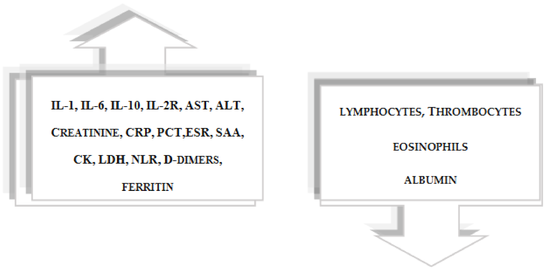
The severity of COVID-19 is correlated with a high level of interleukins IL-6 and IL-1 and with CRP, D-dimers, aspartate aminotransferase (AST), alanine aminotransferase (ALT), creatine kinase (CK), lactate dehydrogenase (LDH), creatinine, low albumin, high erythrocyte sedimentation rate (ESR), low eosinophils, thrombocytopenia and lymphopenia [95,96]. Elevated LDH levels are associated with cell damage. In approximately 80% of patients, lymphopenia and an increased ratio of neutrophils to lymphocytes are observed [16,18,97,98]. Mahat et al. in their review and meta-analysis concluded that in patients with severe COVID-19, serum levels of CRP, ESR, PCT (procalcitonin), IL-6, IL-10, IL-2R, ferritin, SAA (serum amyloid A) and NLR (neutrophil-to-lymphocyte ratio) are significantly increased compared with people with a mild COVID-19. Moreover, they showed increased levels of CRP, PCT, IL-6, ferritin and NLR in non-survivors compared with survivors [99]. Elevated levels of interleukin 6 are characteristic of COVID-19 patients with poor outcomes and are one of the best laboratory indicators of respiratory failure and death. [100,101] IL-6 is considered to be the most significant cytokine in COVID-19, and its increased concentration was also detected in the course of SARS and MERS [102,103]. Elevated serum amyloid A (SAA) levels and disturbances in other biochemical parameters relevant to the development of Alzheimer’s disease have been reported among SARS-CoV-2-positive individuals [99,104,105] (Figure 3). Moreover, the presence of antiphospholipid autoantibodies, which contribute to coagulopathy and ischemic changes in the brain, was observed among COVID-19 patients [24,84,106,107].
4.4. The Specificity of Inflammation in COVID-19
SARS-CoV-2 virus induces heterogeneous immune responses [108]. Some have a lack of or mild immune system response, others develop a strong immune response with a cytokine storm and multi-organ damage, including damage to the brain [86,96,109]. The autoimmune response can be induced by a "molecular mimicry" mechanism, whereby autoimmune cells cross-react with autoantigens [15,110,111]. Research suggests that acute SARS-CoV-2 infection causes a subclinical damage accumulation that predisposes to chronic pro-inflammatory disease and may impair the ability to take full advantage of a strong immune system response to infection or trauma. Infected cells persist for up to 6 weeks from the onset of symptoms, and inflammation may persist for weeks after infection has stopped, suggesting an inadequate, excessive inflammatory response from COVID-19, contributing to multiple organ failure [112]. In response to many stressors, such as oxidative stress, metabolic derangement, altered proteostasis, genome instability, macromolecular damage and many others, cell senescence accumulates, which is involved in the pathogenesis of sustained inflammation of the organism [18]. There is a hypothesis that SARS-CoV-2 may influence cellular ageing by causing excessive oxidative stress, DNA damage and metabolic derangement, as well as resulting in a direct effect of viral infection on tissue damage with activation of inflammation [18]. Additionally, overlapping bacterial infections can exhaust the immune response, with physiological dysregulation contributing to immunoageing [18]. As a result of SARS-CoV-2 infection, innate and adaptive immunity may be exhausted. A hallmark of cellular ageing is the release into the blood of senescence-associated secretory phenotype (SASP) factors, such as cytokines and chemokines, which sustain inflammation [113]. Persistent systemic inflammation due to tissue damage, environmental stressors and psychological and social stresses all influence the risk of developing many chronic diseases, including autoimmune diseases, depression, neurodegenerative disorders, accelerated cognitive decline and dementia [107,114].
4.4.1. Neuroinflammation in COVID-19
The immune system constantly communicates with the brain and spinal cord. Even in the absence of damage to the blood–brain barrier, the T- and B-lymphocytes effectors can penetrate the CNS to destroy pathogens [86,109,115,116]. Neuroinflammation is common in various CNS disease states and is characterised by microglia activation accompanied by leukocyte infiltration [117]. Microglia is highly sensitive to changes in peripheral metabolism and comorbidities as well as to external environmental influences, such as stress, diet, physical activity and environmental pollution [107,118]. Glial cells function as resident cells of the innate immune system response to clearing pathogens and damaged brain tissue, and astrocytes play a role in regulating microcirculation in the brain and regulating extracellular glutamate levels [119]. Astrocytes and microglial cells are responsible for maintaining homeostasis, including neurogenesis, synaptic formation, blood–brain barrier control, capture of reactive oxygen species, neurotransmitter uptake, ion transport and blood flow regulation [107,120,121]. Inflammation may have neurogenesis-regulating properties depending on duration and activity of microglia, astrocytes and macrophages. Proteins CD 47, CD 55 and CD 200 as well as interleukin 4 and 10 secreted by stimulated microglia have proneurogenic properties [122]. However, long-term inflammation, through the activation of microglia, increases the concentration of interleukins IL-6, IL-1β and IL-1α and TNF that are unfavourable for neurogenesis [48,123]. As a result of SARS-CoV-2 infection, brain cells, including neurons, oligodendrocytes and glial cells, may lose their physiological functions, leading to a disturbance of homeostasis in the brain. Even after a SARS-CoV-2 infection has ceased, microglia may remain excited indirectly through epigenetic changes [107,124]. Microglial dysfunction, including disorders of neuronal plasticity, synaptic function, myelination and the blood–brain barrier (BBB) maintenance, can severely impair cognitive function, which may have consequences in the short-term and long-term neuropsychiatric consequences of COVID-19 [125]. It is concluded that disturbances at the cellular and molecular level of microglia and other brain cells, along with the accompanying pandemic stress and anxiety, may contribute to the disclosure of mental disorders, including depressive disorders, psychosis and cognitive disorders such as Alzheimer’s disease, Parkinson’s disease and dementia [20,30,107].
4.4.2. Routes of Infection
There are several routes of infection of the nervous system with SARS-CoV-2. The SARS-CoV-2 virus can infect directly by binding to the ACE2 receptor on endothelial [126], nerve and glial cells [127,128,129,130]. In addition, it can penetrate the central nervous system (CNS) through transnasal and transsynaptic invasions [131,132] to the olfactory bulb and then the brainstem, where it can damage the respiratory centres [107,132].
4.4.3. The Effects of Neuroinflammation
As a result of systemic inflammation, circulating cytokines and chemokines can damage the blood–brain barrier and then the brain parenchyma, leading to neuroinflammation with haemorrhage, leukocyte infiltration and neurodegeneration [19,20]. In severe COVID cases, an increase in blood proinflammatory factors such as a “cytokine storm” results in an effect on the CNS and the manifestation of neuropsychiatric symptoms. [23,28,90,133,134]. Nervous system damage manifests itself through the neurological symptoms of COVID-19, such as acute ischemic stroke, meningitis, encephalopathy and Guillain-Barre syndrome as well as psychiatric disorders, including depressive disorders, delirium and psychosis [12,13,30,135]. A number of neuropsychiatric symptoms are observed in the course of COVID-19, and research also shows the occurrence of neurological and psychiatric complications after the time of infection [136]. The studies conducted so far show the occurrence of symptoms of disturbances of consciousness and deterioration of mental state in approximately 20–30% of patients with severe COVID-19 using neuroimaging with MRI [96,137]. Moreover, in one-third of patients with acute/subacute COVID-19 referred for brain imaging, the results showed hypodense/hyperintense areas on MRI/CT and other abnormalities, such as haemorrhagic lesions, including infarcts [138]. In the study by Coolen et al. postmortem magnetic resonance imaging (MRI) results in non-survivor COVID-19 patients showed in approximately 20% olfactory bulb asymmetry and brain parenchyma abnormalities, including micro- and macro-bleeding and oedema changes [139]. An autopsy study of 21 patients with fatal COVID-19 also revealed extensive inflammation in multiple organs, with no SARS-CoV-2 present in any affected organs. In the examined brain tissue, extensive neutrophilic infiltrates with aggregates of NETs (neutrophil extracellular traps) and platelets were found, without the presence of a virus. The mainly affected areas included the olfactory bulb and medulla oblongata, which explains the symptoms of anosmia and the possible increase in hypoxia with respiratory failure during the disease [112].
5. The COVID-19 Pandemic as a Psychological Stress Factor
The global transmission of the SARS-CoV-2 virus, the lack of reliable information and disinformation fuelled by sensational headlines in the media heightened fears and social phobias [8,9,140]. A state of nationwide quarantine and additional restrictions were introduced, limiting interpersonal contacts and movement [7,141]. There was fear of uncertainty about the course of COVID-19 disease, the timing of the vaccine, the health service’s ability to fight COVID-19 and government procedures to stop the virus from spreading. In addition, there was a strong stigmatisation of people infected with COVID-19 who unknowingly transmitted the infection to other people, observing symptoms of acute stress disorder, self-destructive behaviour and suicide among them [142,143]. There was widespread anxiety and other psychological problems, including the stigmatisation of people infected with the coronavirus in society, especially among medical staff [144,145]. A survey of 2200 Americans, at a time when the number of confirmed COVID-19 cases in the US was 5, with no fatalities, found that 37% of those polled were worried and concerned about the new SARS-CoV-2 virus, and 25% were more concerned in connection with the COVID-19 pandemic than the Ebola outbreak in 2004 [7]. With the exception of the immediate threat of COVID-19, the psychological impact of the pandemic was observed on the mental health of the general population, including tendencies to closely observe the functioning of the body—analysing cough, shortness of breath and constantly monitoring body temperature—and the appearance of mental disorders with a state of anxiety and panic and obsessive-compulsive disorders in connection with justified recommendations for washing and disinfecting hands [146,147,148]. Psychotic exacerbations or even psychosis have been reported among previously mentally unstable people who succumbed to disinformation during the pandemic [149,150]. The impact of strong anxiety accompanying the pandemic on the aggravation of the pre-existing or the emergence of new mental disorders was observed in susceptible people, deprived of natural defence mechanisms due to the lack of social support and suffering from other mental and somatic disorders and previous traumas, without access to reliable information [142,151,152,153]. Nonetheless, psychiatric disorders are a risk with severe COVID-19. A systematic review of studies, including 43,938 COVID-19 patients with comorbid psychiatric disorders, found that the presence of any psychiatric disorder (especially psychotic disorders and mood disorders) and exposure to anti-anxiety and antipsychotic medications were associated with an increased risk of hospitalisation and a higher risk of COVID-19 mortality. This association also applied, to a lesser extent, to intellectual disability, developmental disorders and substance use disorders, but not to anxiety disorders [154].
Furthermore, the impact of the COVID-19 pandemic and its associated social restrictions has been observed in the progression of cognitive impairment in people with dementia and in the exacerbation of neuropsychiatric symptoms in people with chronic neurological diseases [155]. One study examining the effects of quarantine in patients with Alzheimer’s disease (AD), frontotemporal dementia (FTD), vascular dementia (VD) and dementia with Lewy bodies (DLB) found that approximately 60% of patients experienced changes in behavioural and psychological symptoms (BPSD) after one month of isolation, including worsening of previous symptoms (51.9%) or emergence of new symptoms (26%). The most commonly reported symptoms that worsened were irritability, agitation, apathy, depression, anxiety and sleep disturbances. Depending on the type of dementia, severity of prevalence and gender of patients, differences in the trend of reported symptoms were observed. Symptoms of anxiety and depression were more typical for the female gender, patients with AD and mild to moderate disease course. In addition, DLB was associated with a higher risk of increased sleep disturbances and hallucinations as well as FTD with changes in appetite [156]. Another cross-sectional case-control study to quantify anxiety in Parkinson’s disease (PD) patients in relation to social distance restrictions compared with the general population showed the highest levels of anxiety in the PD group [157]. It is suggested that anxiety in PD patients is strongly correlated with the risk of a COVID-19 infection, the availability of the drug in a blockade situation and the perceived higher risk of disease due to chronic comorbidity [157]. In addition, a COVID-19 infection may contribute to the progression of neurodegenerative diseases, such as amyotrophic lateral sclerosis, by exacerbating inflammation, and the progression of the disease state may negatively affect patients’ psychological status. [158]. Outbreaks of pandemics were invariably associated with panic states and a sense of threat to individual safety due to high mortality. Widespread health effects of the pandemic have been reported, including anxiety, insomnia, increased alcohol consumption and loss of energy [159]. Studies on the observed psychological reactions of society during previous pandemics suggest that the response to stress may be influenced by individually diverse factors, such as individual intolerance to uncertainty, perception of one’s own susceptibility to illness and tendency to anxiety [142,160]. One study found that the "vulnerable group" (quarantined or relatives or suspected/ill persons) experienced depressive symptoms more frequently during the SARS pandemic compared with the "non-exposed group". Other studies have found that more than 40% of people who developed SARS experienced symptoms of post-traumatic stress disorder (PTSD) at some point during the pandemic. The respondents who were isolated worked in places with a high risk of SARS infection—e.g., individuals in infectious wards or relatives of patients who experienced contact with SARS were 2–3 times more predisposed to developing severe PTSD symptoms compared with people not exposed to the virus [161]. The above studies and the psychological impact of previous pandemics demonstrate the possible impact of the larger global COVID-19 pandemic on the increase in the incidence of PTSD in the population [162,163,164].
6. Depressive and Neurocognitive Disorders during a COVID-19 Pandemic
The outbreak of a pandemic and rapid changes in almost every aspect of life, social isolation, quarantines and uncertainty about the future can be treated as strong stressors dysfunctionally affecting mental health [165,166]. Studies have shown that COVID-19 patients are at high risk of stress and feelings of stigma [167,168] and are more likely than the general population to suffer from a variety of mental disorders, including depression, anxiety disorders, acute stress disorder, post-traumatic symptoms and insomnia [26,169]. An analysis of COVID-19 patients showed that 1 in 3 presented executive dysfunction and problems with attention and orientation, and areas of hypoperfusion were observed in brain imaging studies [170]. A study of survivors of COVID-19 conducted in Wuhan 6 months after recovery showed symptoms of depression among 23% of patients (367 out of 1617) [171]. One of the prospective studies concerned patients with SARS-CoV-2 admitted to the non-intensive ward, who during hospitalisation showed symptoms of depression in 29% of the patients, while 2 weeks after discharge, in 20% of the patients, symptoms of depression were still observed. This may indicate a relatively stable severity of depression in the subjects shortly after being infected with COVID-19 [172]. Moreover, one of the studies showed that approximately 1 month after infection 31–38% of patients reported symptoms of depression [173]. Another prospective study assessing the mental state and the level of inflammatory markers in COVID-19 survivors one month and three months after discharge from the hospital showed a relationship between inflammation after COVID-19 and depression and related neurocognitive disorders. The 3-month follow-up cohort showed disorders in at least one psychopathological sphere in 35.8% of respondents. Moreover, there was a tendency to intensify depressive symptoms, especially in people with a previous history of mental disorders and in the females, as well as an association of acute infection with the manifestation of neurocognitive disorders including motor coordination and executive dysfunctions in up to 78% of the respondents. With observation, symptoms of depression persisted in contrast with symptoms of anxiety, PTSD and sleep disorders, which improved over time [19]. COVID-19 may develop prolonged inflammation, predisposing to persistent depression and related neurocognitive disorders [18,20,22,24,174]. It is associated with the SARS-CoV-2 infection itself inducing a specific inflammatory response with possible long-term elevated levels of inflammatory markers [18,19,175,176]. In a study from Ireland, approximately 25.3% of 150 patients with COVID-19 had elevated D-dimers 4 months after diagnosis, which was more common in patients requiring hospitalisation and those who were over 50 years of age [177]. A previous study of COVID-19 survivors indicates a relationship between cognitive dysfunction in maintaining attention and underlying inflammation as measured by CRP [125]. Moreover, the baseline level of SI (II) systemic immune inflammation associated with the level of lymphocytes, neutrophils and platelets was found to be related to the severity of depression symptoms and the prediction of neurocognitive impairments [19,178]. In a study by Zhou et al., higher levels of SI (II), which can be considered a marker of the low-grade inflammation observed in a mood disorder [23], have been associated with a major depressive disorder [179]. In one meta-analysis presenting results mainly from China, up to 45% of COVID-19 patients were shown to experience depression, with no gender differences found. However, other studies show that women, people with a severe history of COVID-19, those with elevated markers of inflammation, people with infected family members and patients with an earlier psychiatric diagnosis are in the risk group of developing depressive disorders [178,180].
Post-COVID Syndrome
Symptoms following COVID-19 infection may persist for up to several weeks after an acute infection. Patients with symptoms that persist more than 3 weeks after diagnosis are said to have post-COVID syndrome [181,182]. The syndrome has been suggested to result from prolonged inflammation following a SARS-CoV-2 infection, although the pathogenesis is still under investigation and is not entirely clear. Characteristic symptoms of this syndrome include dyspnoea, chest pain, myalgia, fatigue, and taste and smell disturbances, as well as mental status disorders (Figure 4) [176,183,184].
One study identified the top three symptoms of post-COVID syndrome, which included fatigue, cognitive dysfunction and general malaise [185]. The prospective observational study of patients hospitalised with COVID-19 showed that 4 months after discharge, 51% (244 of 478) had at least one symptom that was not present before the illness, including (31%) reported fatigue, (21%) cognitive symptoms and dyspnoea [184]. In non-hospitalised patients after COVID-19, without comorbidities, it is estimated that 10% to 35% may present with symptoms of post-COVID syndrome [183,186,187], and in hospitalised patients with severe COVID-19, the incidence is up to 80% [182,188,189]. An Italian study of 238 patients hospitalised with severe COVID-19, 4 months after discharge, showed prolonged pulmonary dysfunction in 53.8%, which may account for many post-COVID symptoms [190]. It seems important to follow up on patients, especially in the area of cognitive function, where neurological and cerebrovascular complications were observed during the course of the disease [19,191,192,193]. Direct infection of endothelial cells by SARS-CoV-2 as well as general inflammation contribute to coagulopathy and embolic/thrombotic complications, microcirculatory disorders affecting hypoxia and other symptoms in post-COVID syndrome [136,182,193]. Neurological and cerebrovascular complications are characteristic, although they are less common and may be implicated in the development of Alzheimer’s disease [194]. In addition, it was indicated that in all patients after COVID-19, compared with controls, protein markers of neuronal dysfunction such as amyloid beta, total tau and p-T181, neurogranin and neurofilament light protein were increased in neuron-enriched extracellular vesicles (nEV), indicating an association with neuroinflammation and neurodegeneration [195]. Furthermore, the genetic risk factor of Alzheimer’s disease (ApoE4) has been shown to increase the risk of severe COVID-19 infection, but it is still unclear [196]. It has been reported that 23 of 27 patients with Parkinson’s disease and COVID-99 developed post-COVID syndrome, deterioration of motor function, increased fatigability and increased need for levodopa, as well as sleep and cognitive dysfunction [197]. In one prospective UK study, at least 4 weeks after recovery from acute COVID-19 infection, MRI in more than 70% of patients showed dysfunction in at least one organ, indicating a physiological basis for the infection and possible long-term impairment of body organs [198]. Organ failure was related to heart (systolic dysfunction, myocarditis), liver (hepatomegaly, inflammation, ectopic fat), lung (decreased vital capacity), kidney (inflammation), pancreas (inflammation, ectopic fat) and spleen (splenomegaly). Furthermore, of the incidental more severe structural MRI lesions (n = 56), three were cardiac lesions, and one was renal (hydronephrosis) [198]. Brain MRI findings of a 56-year-old previously healthy patient with neurological symptoms and depression almost 6 months after a COVID-19 infection showed multiple hyperintense areas in the white matter and semi left centres, indicating neurodegeneration and micro-vascular damage [199]. One study of plasma from patients after COVID-19 at 1–3 months found elevated levels of IL-4, which may indicate ongoing neuroinflammation [195]. Another study of 12 patients with neurological complications presenting with post-COVID-19 symptoms between 9 and 12 weeks post-onset found an acute inflammatory phase with significantly elevated inflammatory parameters including C-reactive protein CRP and IL-6 levels [30]. Elevated inflammatory markers may, by impairing the blood–brain barrier, contribute to neuropsychiatric complications including neurocognitive complications through alterations in neurotransmission, including gamma-aminobutyric acid (GABA) [200]. Previous studies have shown in animal models that inflammation with elevated IL-6 levels can decrease GABA receptor density [200]. Furthermore, compared with healthy individuals, evidence of altered neuronal function, neuromotor fatigue, impaired cognitive control, executive dysfunction and apathy was found in patients with post-COVID syndrome [201]. Mental status disorders, including depressive disorders, may affect 26% to 40% of patients even up to 6 months after the onset of symptoms [171,174]. In addition, based on two cases of patients with depression after COVID-19, an association between depression and interleukins, including IL-6, was demonstrated independent of other factors, which may justify the administration of cytokine-reducing drugs to prevent depression after COVID-19 [175]. Moreover, 8 patients with neurological symptoms, compared with 16 without neurological symptoms, had higher levels of anti-SARS-CoV-2 antibodies with an increase in IL-6 [176].
7. Antidepressant Treatment and COVID-19
Depression is associated with low-grade chronic inflammation and is comparable with a chronic cold [44,202]. Patients with chronic inflammatory processes and autoimmune diseases are more prone to depression [43,54,60]. In addition, the assessment of the concentrations of small molecules—metabolites that are products of changes taking place in the body—allows indirect conclusions to be made about the disturbances of specific metabolic pathways [34,203]. The antidepressant treatment used in depressive disorders has antioxidant and anti-inflammatory properties, resulting in a reduction in inflammatory cytokines including CRP and IL-6, [204], with an increase in the concentration of anti-inflammatory cytokines [205,206]. This is supported by neuroimaging studies using PET, showing less neuroinflammation in treated depressed patients compared with untreated patients, which may suggest the normalisation of TSPO expression in the brain and inhibition of microglia activity as a result of antidepressant treatment [73,207]. One study demonstrated a significant effect of antidepressant treatment as well as its duration on the total volume distribution of TSPO in patients with a history of major depressive disorder. In patients untreated for major depressive disorder for 10 years or more, TSPO volume distribution was up to 33% greater in the anterior cingulate cortex, prefrontal cortex and insula compared with participants untreated 9 years or less [208]. It is expected that, in people with a history of COVID-19, the concentration of inflammatory parameters may be higher and the response to antidepressant treatment less effective due to the possible prolonged, abnormal inflammation, with lesions at the level of the nervous system following SARS-CoV-2 infection. Differences in cytokine concentrations in patients may predict disease or resistance to treatment. Based on the current medical knowledge, it is suggested that a history of a SARS-CoV-2 infection may have a significant impact on the reduction in neurocognitive functions [19,125,174,194]. Due to its anti-inflammatory properties, anti-depressant treatment may reduce the inflammatory parameters [209,210]. However, the use of antidepressants to improve neurocognitive impairment is questionable. There are studies indicating the progression of cognitive dysfunction after the inclusion of antidepressants [211,212]. Previous knowledge indicates that, among the selective serotonin reuptake inhibitors (SSRIs) group antidepressants, fluoxetine is a promising drug against COVID-19 through its effect on reducing the secretion of inflammatory cytokines/chemokines (IL-6,CCL-2,TNF-α) and immunomodulatory properties [32,209,210]. In addition, fluoxetine exhibits antiviral activity and is effective against SARS-CoV-2 in cell cultures [32]. This indicates the possible alleviation of neuropsychiatric complications after COVID-19 by fluoxetine use [32]. Such a potent inhibitory effect in both pseudovirus and authentic virus assay has also been described for vortioxetine [213].
8. Discussion
In this review, we present the current knowledge regarding the possible manifestation of depressive and neurocognitive disorders due to the inflammatory background of COVID-19. The experience of previous pandemics, including SARS-CoV, MERS, influenza of the 18th and 19th centuries and Spanish flu in the 20th century, indicates that there is an impact on the development of mental disorders with cognitive deterioration even months after the onset of the disease [18,214]. Studies of convalescents after infection with SARS-CoV and MERS coronaviruses indicate that up to 15% of respondents reported memory, attention and concentration disorders lasting from 6 weeks to 39 months from the onset of the disease [28,29].
Increasingly more studies confirm the hypothesis that inflammation caused by SARS-CoV-2 infection may, in the short term [11,19,178] and long term [2,215], have negative health consequences [182,199,216], including in the field of mental health disorders [18,148,169]. Patients who are severely affected by COVID-19 with respiratory symptoms leaving intensive care units are potentially more likely to experience long-term neuropsychiatric and neurocognitive conditions, such as depression, obsessive-compulsive disorder, psychosis and Parkinson’s and Alzheimer’s disease [20,30]. Studies show the correlation of inflammation with the severity of complications of post-COVID-19 syndrome, including neuropsychiatric disorders [20,30]. Studies of convalescents one month and 3 months after discharge from the hospital indicate an increase in the incidence of psychiatric disorders, including depressive disorders and a deterioration of cognitive functions [28,30,105,217]. In addition, stress during a pandemic, associated with a change in life in almost every aspect, fear of illness, death and other psychosocial factors [7,49,165], can activate the stress axis [46], affect the severity of inflammation in the brain and, consequently, its structural and functional changes [55,65,71,218]. Research confirms that females, the healthcare professionals, elderly individuals, children, college students and psychiatric patients are in the group at increased risk of developing depressive disorders during the COVID-19 pandemic [151,219,220,221,222,223].
An increasing number of studies of COVID-19 survivors show elevated inflammatory markers, including interleukins and CRP, indicating persistent inflammation in the body months after infection [30,175,176,184]. An interesting study found elevated parameters of D-dimers and fibrin products that may be increased during ongoing inflammation and which were observed 6 months after infection with COVID-19 [177]. In addition, observed multisystem inflammatory syndrome in children and adolescents (MIS-C) infection with elevated levels of immunoglobulins, C-reactive protein, ferritin and interleukin-6 indicate possible 4-week post-viral immunisation in the body [224]. Moreover, several cases of adults with MIS-A (multisystem inflammatory syndrome in adults), characterised by a wide spectrum of gastrointestinal, cardiovascular, dermatologic and neurologic symptoms, have been described [225]. As a result of excessive, uncontrolled inflammatory response, immune system dysfunction, including autoimmunity, may occur [18,111]. The presence of antiphospholipid antibodies has been demonstrated in patients with COVID-19, which indicates possible autoimmunity and subsequent attack of the body’s own cells and a number of other complications, including a tendency to thrombosis [106,107].
The analysis of COVID-19 patients showed that every one-third of them presented with executive dysfunction with attention and orientation problems, and areas of hypoperfusion were observed in brain imaging studies [170]. There is evidence that brain hypoperfusion may accelerate amyloid-β (Aβ) accumulation, the pathology of the tau and TDP-43 proteins involved in the development of dementia [105,194]. The white matter of the brain is also very sensitive to ischemia in COVID-19, which affects cognition [194].
The SARS-CoV-2 virus enters the cell by binding ACE2, which is widely expressed on the surface of glial cells, nerve cells and endothelial cells [107]. That is why we observe a number of neuropsychiatric symptoms [12,13,137,150]. Moreover, microglia may remain in an activated state even after cessation of the infection, causing neurotransmission disorders and structural changes in the brain affecting the manifestation of neurocognitive and depressive disorders over time [47,118,120,124,218,226,227]. Although some studies do not indicate the presence of virus in the brain in autopsy studies, the existing neuroinflammation may be the result of a strong systemic inflammatory response damaging the blood–brain barrier [112].
It has been suggested that the COVID-19-related cause of death may be induced by a specific “cytokine storm” with an elevated level of cytokines, (especially IL-1 and IL-6, systemic inflammatory response syndrome—SIRS) leading to multiple organ failure with high mortality [14,15,16,84,86,93]. Recent publications have shown that ACE2 expression is higher in males, which is associated with greater susceptibility to SARS-CoV-2 infection compared with females, which also explains the higher male morbidity and mortality rates [83]. The administration of interleukin 1 and 6 inhibitors in seriously ill COVID-19 patients with respiratory failure and hyperinflammation causes a significant reduction in mortality in patients with IL-1 inhibition. Interleukin 6 inhibition was effective in patients with high levels of CRP, and inhibitions of both IL-1 and IL-6 were effective in patients with low levels of lactate dehydrogenase (LDH) [228].
There are reports of improvement in post-COVID-19 symptoms due to a vaccine that may improve the immune response or reverse autoimmunity [182,229]. In addition, scientific evidence suggests the efficacy of antidepressant treatment having antiviral and anti-inflammatory properties, especially from the SSRI group of drugs, including fluoxetine [32,209,210,213].
Current evidence suggests that symptoms in post-COVID syndrome improve over time and patients show a good prognosis without further sequelae [230]. However, the duration and long-term effects of post-COVID syndrome are unknown and still need further study [31]. It seems important to observe patients, especially in terms of cognitive functions, in whom neurological and cerebrovascular complications were observed during the disease [138,231,232]. There is still insufficient research to establish the exact relationship between the long-term consequences of COVID-19 and the inflammation it causes [18,19,24,84,233].
9. Conclusions
It is increasingly known that, during SARS-CoV-2 infection and after recovery, patients are more likely to develop psychiatric disorders, including depressive and neurocognitive disorders. Further research is required to expand the knowledge on the impact of SARS-CoV-2 infection on the intensification or disclosure of depressive disorders and neurocognitive disorders. Research will broaden our understanding of the possible long-term neuropsychiatric consequences of a COVID-19 infection. At the same time, the studies will help to identify risk groups in terms of difficulties in treating mood disorders, as well as helping to develop potential new therapeutic methods in the future. In addition, the results of the research may increase public awareness of the serious impact of the COVID-19 pandemic on mental health, while paying attention to the need to prevent depression as a serious, potentially life-threatening disease entity. Moreover, it is predicted that, in the coming months and even years, numerous post-COVID-19 patients will seek medical attention from specialists due to complications and symptoms of post-COVID syndrome. It is important to clarify the pathogenesis of SARS-CoV-2 infection and its consequences in the body, including post-COVID-19 syndrome, and to identify markers and targeted therapy. New guidelines are needed for the diagnosis and treatment of this new clinical entity.
10. Limitations
The review did not address the aspect of the manifestation of depressive disorders during COVID-19 in specific age groups, including young adults and the elderly as well as patients with pre-existing inflammatory process. Due to the growing number of studies, only the individual and subjectively most significant results were presented to substantiate the association of inflammation after COVID-19 infection with the manifestation of neurocognitive and depressive disorders. No clear-cut, definite theories of the effect of SARS-CoV-2 on long-term complications exist and, therefore, time for observation, research and further analysis is needed.
Author Contributions
E.D.: Conceptualisation, resources, methodology, writing original draft, review and editing; B.G.-S.: Supervision and review; N.W.: Supervision and review. All authors have read and agreed to the published version of the manuscript.
Funding
This research was supported by the Medical University of Bialystok, grant number SUB/1/DN/21/001/1147. The financial sponsor played no role in the design, execution, analysis or interpretation of the data.
Institutional Review Board Statement
Not applicable.
Informed Consent Statement
Not applicable.
Data Availability Statement
Data sharing not applicable.
Conflicts of Interest
There are no conflicts of interest to declare.
References
- Mukhtar, S. Psychological health during the coronavirus disease 2019 pandemic outbreak. Int. J. Soc. Psychiatry 2020, 66, 512–516. [Google Scholar] [CrossRef]
- De Lorenzo, R.; Conte, C.; Lanzani, C.; Benedetti, F.; Roveri, L.; Mazza, M.G.; Brioni, E.; Giacalone, G.; Canti, V.; Sofia, V.; et al. Residual clinical damage after COVID-19: A retrospective and prospective observational cohort study. PLoS ONE 2020, 15, e0239570. [Google Scholar] [CrossRef]
- Higgins, V.; Sohaei, D.; Diamandis, E.P.; Prassas, I. COVID-19: From an acute to chronic disease? Potential long-term health consequences. Crit. Rev. Clin. Lab. Sci. 2021, 58, 297–310. [Google Scholar] [CrossRef] [PubMed]
- Zhou, P.; Yang, X.-L.; Wang, X.-G.; Hu, B.; Zhang, L.; Zhang, W.; Si, H.-R.; Zhu, Y.; Li, B.; Huang, C.-L.; et al. A pneumonia outbreak associated with a new coronavirus of probable bat origin. Nature 2020, 579, 270–273. [Google Scholar] [CrossRef] [PubMed] [Green Version]
- Cucinotta, D.; Vanelli, M. WHO Declares COVID-19 a Pandemic. Acta Biomed. 2020, 91, 157–160. [Google Scholar] [CrossRef] [PubMed]
- Coronavirus COVID-19 (2019-nCoV). Available online: https://gisanddata.maps.arcgis.com/apps/dashboards/bda7594740fd40299423467b48e9ecf6 (accessed on 30 August 2021).
- Asmundson, G.J.G.; Taylor, S. Coronaphobia: Fear and the 2019-nCoV outbreak. J. Anxiety Disord. 2020, 70, 1–13. [Google Scholar] [CrossRef]
- Khan, S.; Siddique, R.; Li, H.; Ali, A.; Shereen, M.A.; Bashir, N.; Xue, M. Impact of coronavirus outbreak on psychological health. J. Glob. Health 2020, 10. [Google Scholar] [CrossRef]
- Torales, J.; O’Higgins, M.; Castaldelli-Maia, J.M.; Ventriglio, A. The outbreak of COVID-19 coronavirus and its impact on global mental health. Int. J. Soc. Psychiatry 2020, 66, 317–320. [Google Scholar] [CrossRef] [Green Version]
- Szcześniak, D.; Gładka, A.; Misiak, B.; Cyran, A.; Rymaszewska, J. The SARS-CoV-2 and mental health: From biological mechanisms to social consequences. Prog. Neuropsychopharmacol. Biol. Psychiatry 2021, 104, 110046. [Google Scholar] [CrossRef]
- Leung, T.Y.M.; Chan, A.Y.L.; Chan, E.W.; Chan, V.K.Y.; Chui, C.S.L.; Cowling, B.J.; Gao, L.; Ge, M.Q.; Hung, I.F.N.; Ip, M.S.M.; et al. Short- and potential long-term adverse health outcomes of COVID-19: A rapid review. Emerg. Microbes Infect. 2020, 9, 2190–2199. [Google Scholar] [CrossRef]
- Rogers, J.P.; Chesney, E.; Oliver, D.; Pollak, T.A.; McGuire, P.; Fusar-Poli, P.; Zandi, M.S.; Lewis, G.; David, A.S. Psychiatric and neuropsychiatric presentations associated with severe coronavirus infections: A systematic review and meta-analysis with comparison to the COVID-19 pandemic. Lancet Psychiatry 2020, 7, 611–627. [Google Scholar] [CrossRef]
- Ellul, M.A.; Benjamin, L.; Singh, B.; Lant, S.; Michael, B.D.; Easton, A.; Kneen, R.; Defres, S.; Sejvar, J.; Solomon, T. Neurological associations of COVID-19. Lancet Neurol. 2020, 19, 767–783. [Google Scholar] [CrossRef]
- Flores, G. SARS-CoV-2 (COVID-19) has neurotropic and neuroinvasive properties. Int. J. Clin. Pract. 2021, 75, e13708. [Google Scholar] [CrossRef] [PubMed]
- Gammazza, A.M.; Légaré, S.; Bosco, G.L.; Fucarino, A.; Angileri, F.; Oliveri, M.; Cappello, F. Molecular mimicry in the post-COVID-19 signs and symptoms of neurovegetative disorders? Lancet Microbe 2021, 2, e94. [Google Scholar] [CrossRef]
- Zhang, Z.-L.; Hou, Y.-L.; Li, D.-T.; Li, F.-Z. Laboratory findings of COVID-19: A systematic review and meta-analysis. Scand. J. Clin. Lab. Investig. 2020, 80, 441–447. [Google Scholar] [CrossRef]
- Mehta, P.; McAuley, D.F.; Brown, M.; Sanchez, E.; Tattersall, R.S.; Manson, J.J. HLH Across Speciality Collaboration, UK COVID-19: Consider cytokine storm syndromes and immunosuppression. Lancet 2020, 395, 1033–1034. [Google Scholar] [CrossRef]
- Bektas, A.; Schurman, S.H.; Franceschi, C.; Ferrucci, L. A public health perspective of aging: Do hyper-inflammatory syndromes such as COVID-19, SARS, ARDS, cytokine storm syndrome, and post-ICU syndrome accelerate short- and long-term inflammaging? Immun. Ageing 2020, 17, 23. [Google Scholar] [CrossRef]
- Gennaro, M.M.; Mariagrazia, P.; De Lorenzo, R.; Cristiano, M.; Sara, P.; Roberto, F.; Fabio, C.; Patrizia, R.-Q.; Francesco, B. Persistent psychopathology and neurocognitive impairment in COVID-19 survivors: Effect of inflammatory biomarkers at three-month follow-up. Brain Behav. Immun. 2021, 94, 138–147. [Google Scholar] [CrossRef]
- Fotuhi, M.; Mian, A.; Meysami, S.; Raji, C.A. Neurobiology of COVID-19. J. Alzheimer’s Dis. 2020, 76, 3–19. [Google Scholar] [CrossRef]
- Ritchie, K.; Chan, D.; Watermeyer, T. The cognitive consequences of the COVID-19 epidemic: Collateral damage? Brain Commun. 2020, 2, fcaa069. [Google Scholar] [CrossRef]
- Miller, A.H.; Raison, C.L. The role of inflammation in depression: From evolutionary imperative to modern treatment target. Nat. Rev. Immunol. 2016, 16, 22–34. [Google Scholar] [CrossRef] [Green Version]
- Benedetti, F.; Poletti, S.; Hoogenboezem, T.A.; Mazza, E.; Ambrée, O.; de Wit, H.; Wijkhuijs, A.J.M.; Locatelli, C.; Bollettini, I.; Colombo, C.; et al. Inflammatory cytokines influence measures of white matter integrity in Bipolar Disorder. J. Affect. Disord. 2016, 202, 1–9. [Google Scholar] [CrossRef]
- da Silva Lopes, L.; Silva, R.O.; de Sousa Lima, G.; de Araújo Costa, A.C.; Barros, D.F.; Silva-Néto, R.P. Is there a common pathophysiological mechanism between COVID-19 and depression? Acta Neurol. Belg. 2021, 121, 1117–1122. [Google Scholar] [CrossRef]
- Lam, M.H.-B.; Wing, Y.-K.; Yu, M.W.-M.; Leung, C.-M.; Ma, R.C.W.; Kong, A.P.S.; So, W.Y.; Fong, S.Y.-Y.; Lam, S.-P. Mental morbidities and chronic fatigue in severe acute respiratory syndrome survivors: Long-term follow-up. Arch. Intern. Med. 2009, 169, 2142–2147. [Google Scholar] [CrossRef] [Green Version]
- Liu, D.; Baumeister, R.F.; Zhou, Y. Mental health outcomes of coronavirus infection survivors: A rapid meta-analysis. J. Psychiatr. Res. 2020, 137, 542–553. [Google Scholar] [CrossRef]
- De Sousa Moreira, J.L.; Barbosa, S.M.B.; Vieira, J.G.; Chaves, N.C.B.; Felix, E.B.G.; Feitosa, P.W.G.; da Cruz, I.S.; da Silva, C.G.L.; Neto, M.L.R. The psychiatric and neuropsychiatric repercussions associated with severe infections of COVID-19 and other coronaviruses. Prog. Neuropsychopharmacol. Biol. Psychiatry 2021, 106, 110159. [Google Scholar] [CrossRef]
- Hopkins, R.O.; Weaver, L.K.; Pope, D.; Orme, J.F.; Bigler, E.D.; Larson-LOHR, V. Neuropsychological sequelae and impaired health status in survivors of severe acute respiratory distress syndrome. Am. J. Respir. Crit. Care Med. 1999, 160, 50–56. [Google Scholar] [CrossRef]
- Sheng, B.; Cheng, S.K.W.; Lau, K.K.; Li, H.L.; Chan, E.L.Y. The effects of disease severity, use of corticosteroids and social factors on neuropsychiatric complaints in severe acute respiratory syndrome (SARS) patients at acute and convalescent phases. Eur. Psychiatry 2005, 20, 236–242. [Google Scholar] [CrossRef]
- Ortelli, P.; Ferrazzoli, D.; Sebastianelli, L.; Engl, M.; Romanello, R.; Nardone, R.; Bonini, I.; Koch, G.; Saltuari, L.; Quartarone, A.; et al. Neuropsychological and neurophysiological correlates of fatigue in post-acute patients with neurological manifestations of COVID-19: Insights into a challenging symptom. J. Neurol. Sci. 2021, 420, 117271. [Google Scholar] [CrossRef]
- Yelin, D.; Wirtheim, E.; Vetter, P.; Kalil, A.C.; Bruchfeld, J.; Runold, M.; Guaraldi, G.; Mussini, C.; Gudiol, C.; Pujol, M.; et al. Long-term consequences of COVID-19: Research needs. Lancet Infect. Dis. 2020, 20, 1115–1117. [Google Scholar] [CrossRef]
- Pashaei, Y. Drug repurposing of selective serotonin reuptake inhibitors: Could these drugs help fight COVID-19 and save lives? J. Clin. Neurosci. 2021, 88, 163–172. [Google Scholar] [CrossRef]
- Zhang, W.; Zhao, Y.; Zhang, F.; Wang, Q.; Li, T.; Liu, Z.; Wang, J.; Qin, Y.; Zhang, X.; Yan, X.; et al. The use of anti-inflammatory drugs in the treatment of people with severe coronavirus disease 2019 (COVID-19): The Perspectives of clinical immunologists from China. Clin. Immunol. 2020, 214, 108393. [Google Scholar] [CrossRef]
- Nobis, A.; Zalewski, D.; Waszkiewicz, N. Peripheral Markers of Depression. J. Clin. Med. 2020, 9, 3793. [Google Scholar] [CrossRef]
- Archives of Suicide Research—Depression and Suicidal behaviours. Available online: https://www.priory.com/adsui3.htm (accessed on 30 August 2021).
- Marcus, M.; Yasamy, M.T.; van Ommeren, M.; van Chisholm, D.; Saxena, S. Depression: A Global Public Health Concern; WHO: Geneva, Switzerland, 2012. [Google Scholar] [CrossRef] [Green Version]
- Chand, S.P.; Arif, H. Depression; StatPearls Publishing: Treasure Island, FL, USA, 2021. [Google Scholar]
- Gałecki, P.; Talarowska, M. Neurodevelopmental theory of depression. Prog. Neuropsychopharmacol. Biol. Psychiatry 2018, 80, 267–272. [Google Scholar] [CrossRef]
- Dean, J.; Keshavan, M. The neurobiology of depression: An integrated view. Asian J. Psychiatr. 2017, 27, 101–111. [Google Scholar] [CrossRef]
- Maes, M. Major depression and activation of the inflammatory response system. Adv. Exp. Med. Biol. 1999, 461, 25–46. [Google Scholar] [CrossRef]
- McKinnon, M.C.; Yucel, K.; Nazarov, A.; MacQueen, G.M. A meta-analysis examining clinical predictors of hippocampal volume in patients with major depressive disorder. J. Psychiatry Neurosci. 2009, 34, 41–54. [Google Scholar]
- Kempton, M.J.; Salvador, Z.; Munafò, M.R.; Geddes, J.R.; Simmons, A.; Frangou, S.; Williams, S.C.R. Structural neuroimaging studies in major depressive disorder. Meta-analysis and comparison with bipolar disorder. Arch. Gen. Psychiatry 2011, 68, 675–690. [Google Scholar] [CrossRef] [Green Version]
- Patel, A. Review: The role of inflammation in depression. Psychiatr. Danub. 2013, 25 (Suppl. S2), 216–223. [Google Scholar]
- Gałecki, P.; Talarowska, M. Inflammatory theory of depression. Psychiatr. Pol. 2018, 52, 437–447. [Google Scholar] [CrossRef]
- Gałecki, P.; Szulc, A. Psychiatria, 1st ed.; Wydawnictwo Edra Urban & Partner: Wrocław, Poland, 2018; pp. 202–224. [Google Scholar]
- Smith, S.M.; Vale, W.W. The role of the hypothalamic-pituitary-adrenal axis in neuroendocrine responses to stress. Dialogues Clin. Neurosci. 2006, 8, 383–395. [Google Scholar] [PubMed]
- De Pablos, R.M.; Herrera, A.J.; Espinosa-Oliva, A.M.; Sarmiento, M.; Muñoz, M.F.; Machado, A.; Venero, J.L. Chronic stress enhances microglia activation and exacerbates death of nigral dopaminergic neurons under conditions of inflammation. J. Neuroinflamm. 2014, 11, 34. [Google Scholar] [CrossRef] [PubMed] [Green Version]
- Respondek, M.; Buszman, E. Regulacja procesu neurogenezy: Czynniki wpływające na powstawanie nowych komórek nerwowych w mózgu dorosłych ssaków. Postepy Hig. Med. Dosw. 2015, 69, 1451–1461. [Google Scholar]
- Schoenfeld, T.J.; Gould, E. Stress, stress hormones, and adult neurogenesis. Exp. Neurol. 2012, 233, 12–21. [Google Scholar] [CrossRef] [Green Version]
- Vogelzangs, N.; Suthers, K.; Ferrucci, L.; Simonsick, E.M.; Ble, A.; Schrager, M.; Bandinelli, S.; Lauretani, F.; Giannelli, S.V.; Penninx, B.W. Hypercortisolemic depression is associated with the metabolic syndrome in late-life. Psychoneuroendocrinology 2007, 32, 151–159. [Google Scholar] [CrossRef] [Green Version]
- Reus, V.I.; Miner, C. Evidence for physiological effects of hypercortisolemia in psychiatric patients. Psychiatry Res. 1985, 14, 47–56. [Google Scholar] [CrossRef]
- Milne, A.M.B.; MacQueen, G.M.; Hall, G.B.C. Abnormal hippocampal activation in patients with extensive history of major depression: An fMRI study. J. Psychiatry Neurosci. 2012, 37, 28–36. [Google Scholar] [CrossRef] [Green Version]
- Gałecki, P.; Florkowski, A.; Mrowicka, M.; Malinowska, K.; Gałecka, E. Pol. Merkur Lek. 2007, 23, 466–468.
- Troubat, R.; Barone, P.; Leman, S.; Desmidt, T.; Cressant, A.; Atanasova, B.; Brizard, B.; El Hage, W.; Surget, A.; Belzung, C.; et al. Neuroinflammation and depression: A review. Eur. J. Neurosci. 2021, 53, 151–171. [Google Scholar] [CrossRef]
- Maes, M.; Yirmyia, R.; Noraberg, J.; Brene, S.; Hibbeln, J.; Perini, G.; Kubera, M.; Bob, P.; Lerer, B.; Maj, M. The inflammatory & neurodegenerative (I&ND) hypothesis of depression: Leads for future research and new drug developments in depression. Metab. Brain Dis. 2009, 24, 27–53. [Google Scholar] [CrossRef]
- Cerebrospinal Fluid Corticotropin-Releasing Factor (CRF) and Vasopressin Concentrations Predict Pituitary Response in the CRF Stimulation Test: A Multiple Regression Analysis|Neuropsychopharmacology. Available online: https://www.nature.com/articles/1300071 (accessed on 31 August 2021).
- Blume, J.; Douglas, S.D.; Evans, D.L. Immune suppression and immune activation in depression. Brain Behav. Immun. 2011, 25, 221–229. [Google Scholar] [CrossRef] [Green Version]
- Miller, A.H. Depression and immunity: A role for T cells? Brain Behav. Immun. 2010, 24, 1–8. [Google Scholar] [CrossRef] [Green Version]
- Mechanistic Explanations How Cell-Mediated Immune Activation, Inflammation and Oxidative and Nitrosative Stress Pathways and Their Sequels and Concomitants Play a Role in the Pathophysiology of Unipolar Depression. Available online: https://www.sciencedirect.com/science/article/abs/pii/S0149763411002120 (accessed on 31 August 2021).
- Czarny, P.; Wigner, P.; Galecki, P.; Sliwinski, T. The interplay between inflammation, oxidative stress, DNA damage, DNA repair and mitochondrial dysfunction in depression. Prog. Neuropsychopharmacol. Biol. Psychiatry 2018, 80, 309–321. [Google Scholar] [CrossRef]
- Dowlati, Y.; Herrmann, N.; Swardfager, W.; Liu, H.; Sham, L.; Reim, E.K.; Lanctôt, K.L. A meta-analysis of cytokines in major depression. Biol. Psychiatry 2010, 67, 446–457. [Google Scholar] [CrossRef]
- Kim, Y.-K.; Paik, J.-W.; Lee, S.-W.; Yoon, D.; Han, C.; Lee, B.-H. Increased plasma nitric oxide level associated with suicide attempt in depressive patients. Prog. Neuropsychopharmacol. Biol. Psychiatry 2006, 30, 1091–1096. [Google Scholar] [CrossRef]
- Dhir, A.; Kulkarni, S.K. Nitric oxide and major depression. Nitric Oxide 2011, 24, 125–131. [Google Scholar] [CrossRef]
- Lieb, J.; Karmali, R.; Horrobin, D. Elevated levels of prostaglandin E2 and thromboxane B2 in depression. Prostaglandins Leukot. Med. 1983, 10, 361–367. [Google Scholar] [CrossRef]
- Krishnadas, R.; Cavanagh, J. Depression: An inflammatory illness? J. Neurol. Neurosurg. Psychiatry 2012, 83, 495–502. [Google Scholar] [CrossRef] [Green Version]
- Farooq, R.K.; Asghar, K.; Kanwal, S.; Zulqernain, A. Role of inflammatory cytokines in depression: Focus on interleukin-1β. Biomed. Rep. 2017, 6, 15–20. [Google Scholar] [CrossRef] [Green Version]
- Yarlagadda, A.; Alfson, E.; Clayton, A.H. The Blood Brain Barrier and the Role of Cytokines in Neuropsychiatry. Psychiatry 2009, 6, 18–22. [Google Scholar]
- Remus, J.L.; Dantzer, R. Inflammation Models of Depression in Rodents: Relevance to Psychotropic Drug Discovery. Int. J. Neuropsychopharmacol. 2016, 19, 1–13. [Google Scholar] [CrossRef] [PubMed]
- Inflammation and Its Discontents: The Role of Cytokines in the Pathophysiology of Major Depression—ScienceDirect. Available online: https://www.sciencedirect.com/science/article/abs/pii/S0006322308015321 (accessed on 31 August 2021).
- Savitz, J.; Drevets, W.C.; Smith, C.M.; Victor, T.A.; Wurfel, B.E.; Bellgowan, P.S.F.; Bodurka, J.; Teague, T.K.; Dantzer, R. Putative neuroprotective and neurotoxic kynurenine pathway metabolites are associated with hippocampal and amygdalar volumes in subjects with major depressive disorder. Neuropsychopharmacology 2015, 40, 463–471. [Google Scholar] [CrossRef] [PubMed]
- Belleau, E.L.; Treadway, M.T.; Pizzagalli, D.A. The Impact of Stress and Major Depressive Disorder on Hippocampal and Medial Prefrontal Cortex Morphology. Biol. Psychiatry 2019, 85, 443–453. [Google Scholar] [CrossRef] [PubMed]
- Pahwa, R.; Goyal, A.; Bansal, P.; Jialal, I. Chronic Inflammation; StatPearls Publishing: Treasure Island, FL, USA, 2021. [Google Scholar]
- Richards, E.M.; Zanotti-Fregonara, P.; Fujita, M.; Newman, L.; Farmer, C.; Ballard, E.D.; Machado-Vieira, R.; Yuan, P.; Niciu, M.J.; Lyoo, C.H.; et al. PET radioligand binding to translocator protein (TSPO) is increased in unmedicated depressed subjects. EJNMMI Res. 2018, 8, 57. [Google Scholar] [CrossRef]
- Holmes, S.E.; Hinz, R.; Conen, S.; Gregory, C.J.; Matthews, J.C.; Anton-Rodriguez, J.M.; Gerhard, A.; Talbot, P.S. Elevated Translocator Protein in Anterior Cingulate in Major Depression and a Role for Inflammation in Suicidal Thinking: A Positron Emission Tomography Study. Biol. Psychiatry 2018, 83, 61–69. [Google Scholar] [CrossRef] [Green Version]
- Setiawan, E.; Wilson, A.A.; Mizrahi, R.; Rusjan, P.M.; Miler, L.; Rajkowska, G.; Suridjan, I.; Kennedy, J.L.; Rekkas, P.V.; Houle, S.; et al. Role of translocator protein density, a marker of neuroinflammation, in the brain during major depressive episodes. JAMA Psychiatry 2015, 72, 268–275. [Google Scholar] [CrossRef]
- Kokot, F.; Ficek, R. Patogenetyczne i lecznicze aspekty układu reninowo-angiotensynowo-aldosteronowego (RAA)-wczoraj i dziś. Kardiol. Pol. 2001, 54, 295–301. [Google Scholar]
- Atlas, S.A. The renin-angiotensin aldosterone system: Pathophysiological role and pharmacologic inhibition. J. Manag. Care Pharm. 2007, 13, 9–20. [Google Scholar] [CrossRef] [Green Version]
- Hyla-Klekot, L.; Pulcer, B.; Kokot, F. The renin-angiotensin-aldosterone system (RAAS)—New pathogenetic and therapeutic aspects Part. I. Prorenin and renin and its receptors, convertase 2 of angiotensin-1-10, angiotensin-1-7 and its receptor, visceral fat tissue as a site of synthesis of RAAS components. Arter. Hypertens. 2007, 11, 242–247. [Google Scholar]
- Ames, M.K.; Atkins, C.E.; Pitt, B. The renin-angiotensin-aldosterone system and its suppression. J. Vet. Intern Med. 2019, 33, 363–382. [Google Scholar] [CrossRef] [Green Version]
- Gheblawi, M.; Wang, K.; Viveiros, A.; Nguyen, Q.; Zhong, J.-C.; Turner Anthony, J.; Raizada Mohan, K.; Grant Maria, B.; Oudit Gavin, Y. Angiotensin-Converting Enzyme 2: SARS-CoV-2 Receptor and Regulator of the Renin-Angiotensin System. Circ. Res. 2020, 126, 1456–1474. [Google Scholar] [CrossRef]
- Issa, H.; Eid, A.H.; Berry, B.; Takhviji, V.; Khosravi, A.; Mantash, S.; Nehme, R.; Hallal, R.; Karaki, H.; Dhayni, K.; et al. Combination of Angiotensin (1-7) Agonists and Convalescent Plasma as a New Strategy to Overcome Angiotensin Converting Enzyme 2 (ACE2) Inhibition for the Treatment of COVID-19. Front. Med. 2021, 8, 278. [Google Scholar] [CrossRef]
- Heyman, S.N.; Kinaneh, S.; Abassi, Z. The duplicitous nature of ACE2 in COVID-19 disease EBioMedicine (invited commentary). EBioMedicine 2021, 67, 103356. [Google Scholar] [CrossRef]
- Sama, I.E.; Ravera, A.; Santema, B.T.; van Goor, H.; Ter Maaten, J.M.; Cleland, J.G.F.; Rienstra, M.; Friedrich, A.W.; Samani, N.J.; Ng, L.L.; et al. Circulating plasma concentrations of angiotensin-converting enzyme 2 in men and women with heart failure and effects of renin-angiotensin-aldosterone inhibitors. Eur. Heart J. 2020, 41, 1810–1817. [Google Scholar] [CrossRef]
- Tay, M.Z.; Poh, C.M.; Rénia, L.; MacAry, P.A.; Ng, L.F.P. The trinity of COVID-19: Immunity, inflammation and intervention. Nat. Rev. Immunol. 2020, 20, 363–374. [Google Scholar] [CrossRef]
- Bohn, M.K.; Hall, A.; Sepiashvili, L.; Jung, B.; Steele, S.; Adeli, K. Pathophysiology of COVID-19: Mechanisms Underlying Disease Severity and Progression. Physiology 2020, 35, 288–301. [Google Scholar] [CrossRef]
- Huang, C.; Wang, Y.; Li, X.; Ren, L.; Zhao, J.; Hu, Y.; Zhang, L.; Fan, G.; Xu, J.; Gu, X.; et al. Clinical features of patients infected with 2019 novel coronavirus in Wuhan, China. Lancet 2020, 395, 497–506. [Google Scholar] [CrossRef] [Green Version]
- Zhang, B.; Zhou, X.; Qiu, Y.; Song, Y.; Feng, F.; Feng, J.; Song, Q.; Jia, Q.; Wang, J. Clinical characteristics of 82 cases of death from COVID-19. PLoS ONE 2020, 15, e0235458. [Google Scholar] [CrossRef]
- Mahmudpour, M.; Roozbeh, J.; Keshavarz, M.; Farrokhi, S.; Nabipour, I. COVID-19 cytokine storm: The anger of inflammation. Cytokine 2020, 133, 155151. [Google Scholar] [CrossRef]
- Akbari, H.; Tabrizi, R.; Lankarani, K.B.; Aria, H.; Vakili, S.; Asadian, F.; Noroozi, S.; Keshavarz, P.; Faramarz, S. The role of cytokine profile and lymphocyte subsets in the severity of coronavirus disease 2019 (COVID-19): A systematic review and meta-analysis. Life Sci. 2020, 258, 118167. [Google Scholar] [CrossRef]
- Leisman, D.E.; Ronner, L.; Pinotti, R.; Taylor, M.D.; Sinha, P.; Calfee, C.S.; Hirayama, A.V.; Mastroiani, F.; Turtle, C.J.; Harhay, M.O.; et al. Cytokine elevation in severe and critical COVID-19: A rapid systematic review, meta-analysis, and comparison with other inflammatory syndromes. Lancet Respir. Med. 2020, 8, 1233–1244. [Google Scholar] [CrossRef]
- Tanaka, T.; Narazaki, M.; Kishimoto, T. IL-6 in Inflammation, Immunity, and Disease. Cold Spring Harb. Perspect. Biol. 2014, 6, a016295. [Google Scholar] [CrossRef]
- McElvaney, O.J.; Curley, G.F.; Rose-John, S.; McElvaney, N.G. Interleukin-6: Obstacles to targeting a complex cytokine in critical illness. Lancet Respir. Med. 2021, 9, 643–654. [Google Scholar] [CrossRef]
- Jose, R.J.; Manuel, A. COVID-19 cytokine storm: The interplay between inflammation and coagulation. Lancet Respir. Med. 2020, 8, e46–e47. [Google Scholar] [CrossRef]
- de Lucena, T.M.C.; da Silva Santos, A.F.; de Lima, B.R.; de Albuquerque Borborema, M.E.; de Azevêdo Silva, J. Mechanism of inflammatory response in associated comorbidities in COVID-19. Diabetes Metab. Syndr. Clin. Res. Rev. 2020, 14, 597–600. [Google Scholar] [CrossRef]
- Malik, P.; Patel, U.; Mehta, D.; Patel, N.; Kelkar, R.; Akrmah, M.; Gabrilove, J.L.; Sacks, H. Biomarkers and outcomes of COVID-19 hospitalisations: Systematic review and meta-analysis. BMJ Evid. Based Med. 2021, 26, 107–108. [Google Scholar] [CrossRef]
- Chen, T.; Wu, D.; Chen, H.; Yan, W.; Yang, D.; Chen, G. Clinical characteristics of 113 deceased patients with coronavirus disease 2019: Retrospective study. BMJ 2020, 368, m1091. [Google Scholar] [CrossRef] [Green Version]
- Sabbatino, F.; Conti, V.; Franci, G.; Sellitto, C.; Manzo, V.; Pagliano, P.; De Bellis, E.; Masullo, A.; Salzano, F.A.; Caputo, A.; et al. PD-L1 Dysregulation in COVID-19 Patients. Front. Immunol. 2021, 12, 2198. [Google Scholar] [CrossRef] [PubMed]
- Qin, C.; Zhou, L.; Hu, Z.; Zhang, S.; Yang, S.; Tao, Y.; Xie, C.; Ma, K.; Shang, K.; Wang, W.; et al. Dysregulation of Immune Response in Patients with Coronavirus 2019 (COVID-19) in Wuhan, China. Clin. Infect. Dis. 2020, 71, 762–768. [Google Scholar] [CrossRef] [PubMed]
- Mahat, R.K.; Panda, S.; Rathore, V.; Swain, S.; Yadav, L.; Sah, S.P. The dynamics of inflammatory markers in coronavirus disease-2019 (COVID-19) patients: A systematic review and meta-analysis. Clin. Epidemiol. Glob. Health 2021, 100727. [Google Scholar] [CrossRef] [PubMed]
- England, J.T.; Abdulla, A.; Biggs, C.M.; Lee, A.Y.Y.; Hay, K.A.; Hoiland, R.L.; Wellington, C.L.; Sekhon, M.; Jamal, S.; Shojania, K.; et al. Weathering the COVID-19 storm: Lessons from hematologic cytokine syndromes. Blood Rev. 2021, 45, 100707. [Google Scholar] [CrossRef]
- Webb, B.J.; Peltan, I.D.; Jensen, P.; Hoda, D.; Hunter, B.; Silver, A.; Starr, N.; Buckel, W.; Grisel, N.; Hummel, E.; et al. Clinical criteria for COVID-19-associated hyperinflammatory syndrome: A cohort study. Lancet Rheumatol. 2020, 2, e754–e763. [Google Scholar] [CrossRef]
- Costela-Ruiz, V.J.; Illescas-Montes, R.; Puerta-Puerta, J.M.; Ruiz, C.; Melguizo-Rodríguez, L. SARS-CoV-2 infection: The role of cytokines in COVID-19 disease. Cytokine Growth Factor Rev. 2020, 54, 62–75. [Google Scholar] [CrossRef]
- Liu, F.; Li, L.; Xu, M.; Wu, J.; Luo, D.; Zhu, Y.; Li, B.; Song, X.; Zhou, X. Prognostic value of interleukin-6, C-reactive protein, and procalcitonin in patients with COVID-19. J. Clin. Virol. 2020, 127, 104370. [Google Scholar] [CrossRef]
- Shen, X.-N.; Niu, L.-D.; Wang, Y.-J.; Cao, X.-P.; Liu, Q.; Tan, L.; Zhang, C.; Yu, J.-T. Inflammatory markers in Alzheimer’s disease and mild cognitive impairment: A meta-analysis and systematic review of 170 studies. J. Neurol. Neurosurg. Psychiatry 2019, 90, 590–598. [Google Scholar] [CrossRef]
- Rahman, M.A.; Islam, K.; Rahman, S.; Alamin, M. Neurobiochemical Cross-talk Between COVID-19 and Alzheimer’s Disease. Mol. Neurobiol. 2021, 58, 1017–1023. [Google Scholar] [CrossRef]
- Zuo, Y.; Estes, S.K.; Ali, R.A.; Gandhi, A.A.; Yalavarthi, S.; Shi, H.; Sule, G.; Gockman, K.; Madison, J.A.; Zuo, M.; et al. Prothrombotic antiphospholipid antibodies in COVID-19. medRxiv 2020. [Google Scholar] [CrossRef]
- Tremblay, M.-E.; Madore, C.; Bordeleau, M.; Tian, L.; Verkhratsky, A. Neuropathobiology of COVID-19: The Role for Glia. Front. Cell Neurosci. 2020, 14, 592214. [Google Scholar] [CrossRef]
- Desai, N.; Neyaz, A.; Szabolcs, A.; Shih, A.R.; Chen, J.H.; Thapar, V.; Nieman, L.T.; Solovyov, A.; Mehta, A.; Lieb, D.J.; et al. Temporal and spatial heterogeneity of host response to SARS-CoV-2 pulmonary infection. Nat Commun. 2020, 11, 6319. [Google Scholar] [CrossRef]
- Chen, Y.; Li, L. SARS-CoV-2: Virus dynamics and host response. Lancet Infect. Dis. 2020, 20, 515–516. [Google Scholar] [CrossRef] [Green Version]
- Ehrenfeld, M.; Tincani, A.; Andreoli, L.; Cattalini, M.; Greenbaum, A.; Kanduc, D.; Alijotas-Reig, J.; Zinserling, V.; Semenova, N.; Amital, H.; et al. COVID-19 and autoimmunity. Autoimmun. Rev. 2020, 19, 102597. [Google Scholar] [CrossRef]
- Does SARS-CoV-2 Trigger Stress-InducedAutoimmunity by Molecular Mimicry? A Hypothesis. Available online: https://pubmed.ncbi.nlm.nih.gov/32610587/ (accessed on 31 August 2021).
- Schurink, B.; Roos, E.; Radonic, T.; Barbe, E.; Bouman, C.S.C.; de Boer, H.H.; de Bree, G.J.; Bulle, E.B.; Aronica, E.M.; Florquin, S.; et al. Viral presence and immunopathology in patients with lethal COVID-19: A prospective autopsy cohort study. Lancet Microbe 2020, 1, e290–e299. [Google Scholar] [CrossRef]
- Alster, O.; Korwek, Z. Znaczniki starzenia komórkowego. Postepy Biochem. 2014, 60, 138–146. [Google Scholar]
- Sartori, A.C.; Vance, D.E.; Slater, L.Z.; Crowe, M. The Impact of Inflammation on Cognitive Function in Older Adults: Implications for Health Care Practice and Research. J. Neurosci. Nurs. 2012, 44, 206–217. [Google Scholar] [CrossRef] [Green Version]
- Mao, L.; Jin, H.; Wang, M.; Hu, Y.; Chen, S.; He, Q.; Chang, J.; Hong, C.; Zhou, Y.; Wang, D.; et al. Neurologic Manifestations of Hospitalized Patients With Coronavirus Disease 2019 in Wuhan, China. JAMA Neurol. 2020, 77, 683–690. [Google Scholar] [CrossRef] [Green Version]
- Aarli, J.A. The immune system and the nervous system. J. Neurol. 1983, 229, 137–154. [Google Scholar] [CrossRef]
- Schwartz, M.; Deczkowska, A. Neurological Disease as a Failure of Brain-Immune Crosstalk: The Multiple Faces of Neuroinflammation. Trends Immunol. 2016, 37, 668–679. [Google Scholar] [CrossRef]
- Tay, T.L.; Savage, J.C.; Hui, C.W.; Bisht, K.; Tremblay, M.-È. Microglia across the lifespan: From origin to function in brain development, plasticity and cognition. J. Physiol. 2017, 595, 1929–1945. [Google Scholar] [CrossRef] [Green Version]
- Engelhardt, B.; Vajkoczy, P.; Weller, R.O. The movers and shapers in immune privilege of the CNS. Nat. Immunol. 2017, 18, 123–131. [Google Scholar] [CrossRef] [PubMed]
- Hanisch, U.-K.; Kettenmann, H. Microglia: Active sensor and versatile effector cells in the normal and pathologic brain. Nat. Neurosci. 2007, 10, 1387–1394. [Google Scholar] [CrossRef]
- Verkhratsky, A.; Zorec, R.; Parpura, V. Stratification of astrocytes in healthy and diseased brain. Brain Pathol. 2017, 27, 629–644. [Google Scholar] [CrossRef]
- Fuster-Matanzo, A.; Llorens-Martín, M.; Hernández, F.; Avila, J. Role of neuroinflammation in adult neurogenesis and Alzheimer disease: Therapeutic approaches. Mediat. Inflamm. 2013, 2013, 260925. [Google Scholar] [CrossRef] [PubMed] [Green Version]
- Regulation of Adult Neurogenesis by Stress, Sleep Disruption, Exercise and Inflammation: Implications for Depression and Antidepressant Action. Available online: https://www.sciencedirect.com/science/article/pii/S0924977X09002119 (accessed on 1 September 2021).
- Cheray, M.; Joseph, B. Epigenetics Control Microglia Plasticity. Front. Cell Neurosci. 2018, 12, 243. [Google Scholar] [CrossRef] [PubMed] [Green Version]
- Zhou, H.; Lu, S.; Chen, J.; Wei, N.; Wang, D.; Lyu, H.; Shi, C.; Hu, S. The landscape of cognitive function in recovered COVID-19 patients. J. Psychiatr. Res. 2020, 129, 98–102. [Google Scholar] [CrossRef] [PubMed]
- Zeisel, A.; Muñoz-Manchado, A.B.; Codeluppi, S.; Lönnerberg, P.; La Manno, G.; Juréus, A.; Marques, S.; Munguba, H.; He, L.; Betsholtz, C.; et al. Brain structure. Cell types in the mouse cortex and hippocampus revealed by single-cell RNA-seq. Science 2015, 347, 1138–1142. [Google Scholar] [CrossRef] [PubMed]
- Gowrisankar, Y.V.; Clark, M.A. Angiotensin II regulation of angiotensin-converting enzymes in spontaneously hypertensive rat primary astrocyte cultures. J. Neurochem. 2016, 138, 74–85. [Google Scholar] [CrossRef] [PubMed] [Green Version]
- Nemoto, W.; Yamagata, R.; Nakagawasai, O.; Nakagawa, K.; Hung, W.-Y.; Fujita, M.; Tadano, T.; Tan-No, K. Effect of spinal angiotensin-converting enzyme 2 activation on the formalin-induced nociceptive response in mice. Eur. J. Pharmacol. 2020, 872, 172950. [Google Scholar] [CrossRef]
- Tekin, H.; Simmons, S.; Cummings, B.; Gao, L.; Adiconis, X.; Hession, C.C.; Ghoshal, A.; Dionne, D.; Choudhury, S.R.; Yesilyurt, V.; et al. Effects of 3D culturing conditions on the transcriptomic profile of stem-cell-derived neurons. Nat. Biomed. Eng. 2018, 2, 540–554. [Google Scholar] [CrossRef]
- Xia, H.; Lazartigues, E. Angiotensin-Converting Enzyme 2: Central Regulator for Cardiovascular Function. Curr. Hypertens Rep. 2010, 12, 170–175. [Google Scholar] [CrossRef]
- Netland, J.; Meyerholz, D.K.; Moore, S.; Cassell, M.; Perlman, S. Severe acute respiratory syndrome coronavirus infection causes neuronal death in the absence of encephalitis in mice transgenic for human ACE2. J. Virol. 2008, 82, 7264–7275. [Google Scholar] [CrossRef] [Green Version]
- Li, K.; Wohlford-Lenane, C.; Perlman, S.; Zhao, J.; Jewell, A.K.; Reznikov, L.R.; Gibson-Corley, K.N.; Meyerholz, D.K.; McCray, P.B., Jr. Middle East Respiratory Syndrome Coronavirus Causes Multiple Organ Damage and Lethal Disease in Mice Transgenic for Human Dipeptidyl Peptidase 4. J. Infect. Dis. 2016, 213, 712–722. [Google Scholar] [CrossRef] [Green Version]
- De Picker, L.J.; Yolken, R.; Benedetti, F.; Borsini, A.; Branchi, I.; Fusar-Poli, P.; Carlos Leza, J.; Pariante, C.; Pollak, T.; Tamouza, R.; et al. Viewpoint|European COVID-19 exit strategy for people with severe mental disorders: Too little, but not yet too late. Brain Behav. Immun. 2021, 94, 15–178. [Google Scholar] [CrossRef]
- Hu, B.; Huang, S.; Yin, L. The cytokine storm and COVID-19. J. Med. Virol. 2021, 93, 250–256. [Google Scholar] [CrossRef]
- Romero-Sánchez, C.M.; Díaz-Maroto, I.; Fernández-Díaz, E.; Sánchez-Larsen, Á.; Layos-Romero, A.; García-García, J.; González, E.; Redondo-Peñas, I.; Perona-Moratalla, A.B.; Del Valle-Pérez, J.A.; et al. Neurologic manifestations in hospitalized patients with COVID-19: The ALBACOVID registry. Neurology 2020, 95, e1060–e1070. [Google Scholar] [CrossRef]
- Taquet, M.; Geddes, J.R.; Husain, M.; Luciano, S.; Harrison, P.J. 6-month neurological and psychiatric outcomes in 236 379 survivors of COVID-19: A retrospective cohort study using electronic health records. Lancet Psychiatry 2021, 8, 416–427. [Google Scholar] [CrossRef]
- Varatharaj, A.; Thomas, N.; Ellul, M.A.; Davies, N.W.S.; Pollak, T.A.; Tenorio, E.L.; Sultan, M.; Easton, A.; Breen, G.; Zandi, M.; et al. Neurological and neuropsychiatric complications of COVID-19 in 153 patients: A UK-wide surveillance study. Lancet Psychiatry 2020, 7, 875–882. [Google Scholar] [CrossRef]
- Egbert, A.R.; Cankurtaran, S.; Karpiak, S. Brain abnormalities in COVID-19 acute/subacute phase: A rapid systematic review. Brain Behav. Immun. 2020, 89, 543–554. [Google Scholar] [CrossRef]
- Coolen, T.; Lolli, V.; Sadeghi, N.; Rovai, A.; Trotta, N.; Taccone, F.S.; Creteur, J.; Henrard, S.; Goffard, J.-C.; Dewitte, O.; et al. Early postmortem brain MRI findings in COVID-19 non-survivors. Neurology 2020, 95, e2016–e2027. [Google Scholar] [CrossRef]
- Onyeaka, H.K.; Zahid, S.; Patel, R.S. The Unaddressed Behavioral Health Aspect During the Coronavirus Pandemic. Cureus 2020, 12, e7351. [Google Scholar] [CrossRef] [Green Version]
- Brooks, S.K.; Webster, R.K.; Smith, L.E.; Woodland, L.; Wessely, S.; Greenberg, N.; Rubin, G.J. The psychological impact of quarantine and how to reduce it: Rapid review of the evidence. Lancet 2020, 395, 912–920. [Google Scholar] [CrossRef] [Green Version]
- Heitzman, J. Impact of COVID-19 pandemic on mental health. Psychiatr. Pol. 2020, 54, 187–198. [Google Scholar] [CrossRef] [PubMed]
- Liu, Y.; Cao, L.; Li, X.; Jia, Y.; Xia, H. Awareness of mental health problems in patients with coronavirus disease 19 (COVID-19): A lesson from an adult man attempting suicide. Asian J. Psychiatr. 2020, 51, 102106. [Google Scholar] [CrossRef] [PubMed]
- Possible Special Needs for Mental Online Support in Female and Male Health Care Workers during the COVID-19-Waszkiewicz. 2021. Available online: https://onlinelibrary.wiley.com/doi/full/10.1002/jmv.26273 (accessed on 1 September 2021).
- Sorokin, M.Y.; Kasyanov, E.D.; Rukavishnikov, G.V.; Makarevich, O.V.; Neznanov, N.G.; Morozov, P.V.; Lutova, N.B.; Mazo, G.E. Stress and Stigmatization in Health-Care Workers during the COVID-19 Pandemic. Indian J. Psychiatry 2020, 62, S445–S453. [Google Scholar] [CrossRef] [PubMed]
- Jassi, A.; Shahriyarmolki, K.; Taylor, T.; Peile, L.; Challacombe, F.; Clark, B.; Veale, D. OCD and COVID-19: A new frontier. Cogn. Behav. Therap. 2020, 13, e27. [Google Scholar] [CrossRef] [PubMed]
- Dubey, S.; Biswas, P.; Ghosh, R.; Chatterjee, S.; Dubey, M.J.; Chatterjee, S.; Lahiri, D.; Lavie, C.J. Psychosocial impact of COVID-19. Diabetes Metab. Syndr. 2020, 14, 779–788. [Google Scholar] [CrossRef] [PubMed]
- Li, W.; Yang, Y.; Liu, Z.-H.; Zhao, Y.-J.; Zhang, Q.; Zhang, L.; Cheung, T.; Xiang, Y.-T. Progression of Mental Health Services during the COVID-19 Outbreak in China. Int. J. Biol. Sci. 2020, 16, 1732–1738. [Google Scholar] [CrossRef] [Green Version]
- Colizzi, M.; Bortoletto, R.; Silvestri, M.; Mondini, F.; Puttini, E.; Cainelli, C.; Gaudino, R.; Ruggeri, M.; Zoccante, L. Medically unexplained symptoms in the times of COVID-19 pandemic: A case-report. Brain Behav. Immun. Health 2020, 5, 100073. [Google Scholar] [CrossRef]
- Ferrando, S.J.; Klepacz, L.; Lynch, S.; Tavakkoli, M.; Dornbush, R.; Baharani, R.; Smolin, Y.; Bartell, A. COVID-19 Psychosis: A Potential New Neuropsychiatric Condition Triggered by Novel Coronavirus Infection and the Inflammatory Response? Psychosomatics 2020, 61, 551–555. [Google Scholar] [CrossRef]
- PRIME PubMed | Psychological Stress Risk Factors, Concerns and Mental Health Support Among Health Care Workers in Vietnam During the Coronavirus Disease 2019 (COVID-19) Outbreak. Available online: https://brain.unboundmedicine.com/medline/citation/33816419/Psychological_Stress_Risk_Factors,_Concerns_and_Mental_Health_Support_Among_Health_Care_Workers_in_Vietnam_During_the_Coronavirus_Disease_2019_(COVID-19)_Outbreak (accessed on 1 September 2021).
- Vos, L.M.W.; Habibović, M.; Nyklíček, I.; Smeets, T.; Mertens, G. Optimism, mindfulness, and resilience as potential protective factors for the mental health consequences of fear of the coronavirus. Psychiatry Res. 2021, 300, 113927. [Google Scholar] [CrossRef]
- Chatterjee, S.S.; Barikar, C.M.; Mukherjee, A. Impact of COVID-19 pandemic on pre-existing mental health problems. Asian J. Psychiatr. 2020, 51, 102071. [Google Scholar] [CrossRef]
- Vai, B.; Mazza, M.G.; Delli Colli, C.; Foiselle, M.; Allen, B.; Benedetti, F.; Borsini, A.; Casanova Dias, M.; Tamouza, R.; Leboyer, M.; et al. Mental disorders and risk of COVID-19-related mortality, hospitalisation, and intensive care unit admission: A systematic review and meta-analysis. Lancet Psychiatry 2021, 8, 797–812. [Google Scholar] [CrossRef]
- Tondo, G.; Sarasso, B.; Serra, P.; Tesser, F.; Comi, C. The Impact of the COVID-19 Pandemic on the Cognition of People with Dementia. Int. J. Environ. Res. Public Health 2021, 18, 4285. [Google Scholar] [CrossRef]
- Cagnin, A.; Di Lorenzo, R.; Marra, C.; Bonanni, L.; Cupidi, C.; Laganà, V.; Rubino, E.; Vacca, A.; Provero, P.; Isella, V.; et al. Behavioral and Psychological Effects of Coronavirus Disease-19 Quarantine in Patients With Dementia. Front. Psychiatry 2020, 11, 578015. [Google Scholar] [CrossRef]
- Salari, M.; Zali, A.; Ashrafi, F.; Etemadifar, M.; Sharma, S.; Hajizadeh, N.; Ashourizadeh, H. Incidence of Anxiety in Parkinson’s Disease During the Coronavirus Disease (COVID-19) Pandemic. Mov. Disord. 2020, 35, 1095–1096. [Google Scholar] [CrossRef]
- Li, X.; Bedlack, R. COVID-19-accelerated disease progression in two patients with amyotrophic lateral sclerosis. Muscle Nerve 2021, 64, E13–E15. [Google Scholar] [CrossRef]
- Preti, E.; Di Mattei, V.; Perego, G.; Ferrari, F.; Mazzetti, M.; Taranto, P.; Di Pierro, R.; Madeddu, F.; Calati, R. The Psychological Impact of Epidemic and Pandemic Outbreaks on Healthcare Workers: Rapid Review of the Evidence. Curr. Psychiatry Rep. 2020, 22, 43. [Google Scholar] [CrossRef]
- Vindegaard, N.; Benros, M.E. COVID-19 pandemic and mental health consequences: Systematic review of the current evidence. Brain Behav. Immun. 2020, 89, 531–542. [Google Scholar] [CrossRef]
- Liu, N.; Zhang, F.; Wei, C.; Jia, Y.; Shang, Z.; Sun, L.; Wu, L.; Sun, Z.; Zhou, Y.; Wang, Y.; et al. Prevalence and predictors of PTSS during COVID-19 outbreak in China hardest-hit areas: Gender differences matter. Psychiatry Res. 2020, 287, 112921. [Google Scholar] [CrossRef]
- Tarsitani, L.; Vassalini, P.; Koukopoulos, A.; Borrazzo, C.; Alessi, F.; Di Nicolantonio, C.; Serra, R.; Alessandri, F.; Ceccarelli, G.; Mastroianni, C.M.; et al. Post-traumatic Stress Disorder Among COVID-19 Survivors at 3-Month Follow-up After Hospital Discharge. J. Gen. Intern. Med. 2021, 36, 1702–1707. [Google Scholar] [CrossRef]
- Horesh, D.; Brown, A.D. Traumatic stress in the age of COVID-19: A call to close critical gaps and adapt to new realities. Psychol. Trauma 2020, 12, 331–335. [Google Scholar] [CrossRef]
- Survivors of COVID-19 Are at High Risk of Posttraumatic Stress Disorder. Available online: https://pubmed.ncbi.nlm.nih.gov/32514428/ (accessed on 1 September 2021).
- Bridgland, V.M.E.; Moeck, E.K.; Green, D.M.; Swain, T.L.; Nayda, D.M.; Matson, L.A.; Hutchison, N.P.; Takarangi, M.K.T. Why the COVID-19 pandemic is a traumatic stressor. PLoS ONE 2021, 16, e0240146. [Google Scholar] [CrossRef]
- Stanton, R.; To, Q.G.; Khalesi, S.; Williams, S.L.; Alley, S.J.; Thwaite, T.L.; Fenning, A.S.; Vandelanotte, C. Depression, Anxiety and Stress during COVID-19: Associations with Changes in Physical Activity, Sleep, Tobacco and Alcohol Use in Australian Adults. Int. J. Environ. Res. Public Health 2020, 17, 4065. [Google Scholar] [CrossRef]
- Abuhammad, S.; Alzoubi, K.H.; Khabour, O. Fear of COVID-19 and stigmatization towards infected people among Jordanian people. Int. J. Clin. Pract. 2020, e13899. [Google Scholar] [CrossRef]
- Bhanot, D.; Singh, T.; Verma, S.K.; Sharad, S. Stigma and Discrimination during COVID-19 Pandemic. Front. Public Health 2021, 8, 577018. [Google Scholar] [CrossRef] [PubMed]
- Kathirvel, N. Post COVID-19 pandemic mental health challenges. Asian J. Psychiatr. 2020, 53, 102430. [Google Scholar] [CrossRef] [PubMed]
- Helms, J.; Kremer, S.; Merdji, H.; Clere-Jehl, R.; Schenck, M.; Kummerlen, C.; Collange, O.; Boulay, C.; Fafi-Kremer, S.; Ohana, M.; et al. Neurologic Features in Severe SARS-CoV-2 Infection. N. Engl. J. Med. 2020, 382, 2268–2270. [Google Scholar] [CrossRef]
- Huang, C.; Huang, L.; Wang, Y.; Li, X.; Ren, L.; Gu, X.; Kang, L.; Guo, L.; Liu, M.; Zhou, X.; et al. 6-month consequences of COVID-19 in patients discharged from hospital: A cohort study. Lancet 2021, 397, 220–232. [Google Scholar] [CrossRef]
- Parker, C.; Shalev, D.; Hsu, I.; Shenoy, A.; Cheung, S.; Nash, S.; Wiener, I.; Fedoronko, D.; Allen, N.; Shapiro, P.A. Depression, Anxiety, and Acute Stress Disorder Among Patients Hospitalized With COVID-19: A Prospective Cohort Study. J. Acad. Consult. Liaison Psychiatry 2021, 62, 211–219. [Google Scholar] [CrossRef] [PubMed]
- Cai, X.; Hu, X.; Ekumi, I.O.; Wang, J.; An, Y.; Li, Z.; Yuan, B. Psychological Distress and Its Correlates among COVID-19 Survivors During Early Convalescence Across Age Groups. Am. J. Geriatr. Psychiatry 2020, 28, 1030–1039. [Google Scholar] [CrossRef] [PubMed]
- Alemanno, F.; Houdayer, E.; Parma, A.; Spina, A.; Del Forno, A.; Scatolini, A.; Angelone, S.; Brugliera, L.; Tettamanti, A.; Beretta, L.; et al. COVID-19 cognitive deficits after respiratory assistance in the subacute phase: A COVID-rehabilitation unit experience. PLoS ONE 2021, 16, e0246590. [Google Scholar] [CrossRef] [PubMed]
- Alpert, O.; Begun, L.; Garren, P.; Solhkhah, R. Cytokine storm induced new onset depression in patients with COVID-19. A new look into the association between depression and cytokines -two case reports. Brain Behav. Immun. Health 2020, 9, 100173. [Google Scholar] [CrossRef]
- Sun, B.; Tang, N.; Peluso, M.J.; Iyer, N.S.; Torres, L.; Donatelli, J.L.; Munter, S.E.; Nixon, C.C.; Rutishauser, R.L.; Rodriguez-Barraquer, I.; et al. Characterization and Biomarker Analyses of Post-COVID-19 Complications and Neurological Manifestations. Cells 2021, 10, 386. [Google Scholar] [CrossRef]
- Townsend, L.; Fogarty, H.; Dyer, A.; Martin-Loeches, I.; Bannan, C.; Nadarajan, P.; Bergin, C.; O’Farrelly, C.; Conlon, N.; Bourke, N.M.; et al. Prolonged elevation of D-dimer levels in convalescent COVID-19 patients is independent of the acute phase response. J. Thromb Haemost. 2021. [Google Scholar] [CrossRef]
- Mazza, M.G.; De Lorenzo, R.; Conte, C.; Poletti, S.; Vai, B.; Bollettini, I.; Melloni, E.M.T.; Furlan, R.; Ciceri, F.; Rovere-Querini, P.; et al. Anxiety and depression in COVID-19 survivors: Role of inflammatory and clinical predictors. Brain Behav. Immun. 2020, 89, 594–600. [Google Scholar] [CrossRef]
- Zhou, L.; Ma, X.; Wang, W. Inflammation and Coronary Heart Disease Risk in Patients with Depression in China Mainland: A Cross-Sectional Study. Neuropsychiatr. Dis. Treat. 2020, 16, 81–86. [Google Scholar] [CrossRef] [Green Version]
- Ma, Y.-F.; Li, W.; Deng, H.-B.; Wang, L.; Wang, Y.; Wang, P.-H.; Bo, H.-X.; Cao, J.; Wang, Y.; Zhu, L.-Y.; et al. Prevalence of depression and its association with quality of life in clinically stable patients with COVID-19. J. Affect. Disord. 2020, 275, 145–148. [Google Scholar] [CrossRef]
- Amenta, E.M.; Spallone, A.; Rodriguez-Barradas, M.C.; El Sahly, H.M.; Atmar, R.L.; Kulkarni, P.A. Postacute COVID-19: An Overview and Approach to Classification. Open Forum Infect. Dis. 2020, 7, ofaa509. [Google Scholar] [CrossRef]
- Maltezou, H.C.; Pavli, A.; Tsakris, A. Post-COVID Syndrome: An Insight on Its Pathogenesis. Vaccines 2021, 9, 497. [Google Scholar] [CrossRef]
- Havervall, S.; Rosell, A.; Phillipson, M.; Mangsbo, S.M.; Nilsson, P.; Hober, S.; Thålin, C. Symptoms and Functional Impairment Assessed 8 Months After Mild COVID-19 Among Health Care Workers. JAMA 2021, 325, 2015–2016. [Google Scholar] [CrossRef]
- Writing Committee for the COMEBAC Study Group; Morin, L.; Savale, L.; Pham, T.; Colle, R.; Figueiredo, S.; Harrois, A.; Gasnier, M.; Lecoq, A.-L.; Meyrignac, O.; et al. Four-Month Clinical Status of a Cohort of Patients After Hospitalization for COVID-19. JAMA 2021, 325, 1525–1534. [Google Scholar] [CrossRef]
- Davis, H.E.; Assaf, G.S.; McCorkell, L.; Wei, H.; Low, R.J.; Re’em, Y.; Redfield, S.; Austin, J.P.; Akrami, A. Characterizing long COVID in an international cohort: 7 months of symptoms and their impact. eClinicalMedicine 2021, 38, 101019. [Google Scholar] [CrossRef]
- Greenhalgh, T.; Knight, M.; A’Court, C.; Buxton, M.; Husain, L. Management of post-acute COVID-19 in primary care. BMJ 2020, 370, m3026. [Google Scholar] [CrossRef]
- Tenforde, M.W.; Kim, S.S.; Lindsell, C.J.; Billig Rose, E.; Shapiro, N.I.; Files, D.C.; Gibbs, K.W.; Erickson, H.L.; Steingrub, J.S.; Smithline, H.A.; et al. Symptom Duration and Risk Factors for Delayed Return to Usual Health Among Outpatients with COVID-19 in a Multistate Health Care Systems Network—United States, March-June 2020. MMWR Morb. Mortal. Wkly Rep. 2020, 69, 993–998. [Google Scholar] [CrossRef]
- Carvalho-Schneider, C.; Laurent, E.; Lemaignen, A.; Beaufils, E.; Bourbao-Tournois, C.; Laribi, S.; Flament, T.; Ferreira-Maldent, N.; Bruyère, F.; Stefic, K.; et al. Follow-up of adults with noncritical COVID-19 two months after symptom onset. Clin. Microbiol. Infect. 2021, 27, 258–263. [Google Scholar] [CrossRef] [PubMed]
- Becker, R.C. COVID-19 and its sequelae: A platform for optimal patient care, discovery and training. J. Thromb. Thrombolysis 2021, 51, 587–594. [Google Scholar] [CrossRef] [PubMed]
- Bellan, M.; Soddu, D.; Balbo, P.E.; Baricich, A.; Zeppegno, P.; Avanzi, G.C.; Baldon, G.; Bartolomei, G.; Battaglia, M.; Battistini, S.; et al. Respiratory and Psychophysical Sequelae Among Patients With COVID-19 Four Months After Hospital Discharge. JAMA Network Open 2021, 4, e2036142. [Google Scholar] [CrossRef] [PubMed]
- Baig, A.M. Updates on What ACS Reported: Emerging Evidences of COVID-19 with Nervous System Involvement. ACS Chem. Neurosci. 2020, 11, 1204–1205. [Google Scholar] [CrossRef] [PubMed]
- Huang, Y.H.; Jiang, D.; Huang, J.T. SARS-CoV-2 Detected in Cerebrospinal Fluid by PCR in a Case of COVID-19 Encephalitis. Brain Behav. Immun. 2020, 87, 149. [Google Scholar] [CrossRef]
- Lu, Y.; Li, X.; Geng, D.; Mei, N.; Wu, P.-Y.; Huang, C.-C.; Jia, T.; Zhao, Y.; Wang, D.; Xiao, A.; et al. Cerebral Micro-Structural Changes in COVID-19 Patients—An MRI-based 3-month Follow-up Study. eClinicalMedicine 2020, 25. [Google Scholar] [CrossRef]
- Miners, S.; Kehoe, P.G.; Love, S. Cognitive impact of COVID-19: Looking beyond the short term. Alzheimers Res. Ther. 2020, 12, 170. [Google Scholar] [CrossRef]
- Koumpa, F.S.; Forde, C.T.; Manjaly, J.G. Sudden irreversible hearing loss post COVID-19. BMJ Case Rep. 2020, 13, e238419. [Google Scholar] [CrossRef]
- Wang, C.; Zhang, M.; Garcia, G.; Tian, E.; Cui, Q.; Chen, X.; Sun, G.; Wang, J.; Arumugaswami, V.; Shi, Y. ApoE-Isoform-Dependent SARS-CoV-2 Neurotropism and Cellular Response. Cell Stem Cell 2021, 28, 331–342.e5. [Google Scholar] [CrossRef]
- Leta, V.; Rodríguez-Violante, M.; Abundes, A.; Rukavina, K.; Teo, J.T.; Falup-Pecurariu, C.; Irincu, L.; Rota, S.; Bhidayasiri, R.; Storch, A.; et al. Parkinson’s Disease and Post–COVID-19 Syndrome: The Parkinson’s Long-COVID Spectrum. Mov. Disord. 2021, 36, 1287–1289. [Google Scholar] [CrossRef]
- Dennis, A.; Wamil, M.; Alberts, J.; Oben, J.; Cuthbertson, D.J.; Wootton, D.; Crooks, M.; Gabbay, M.; Brady, M.; Hishmeh, L.; et al. Multiorgan impairment in low-risk individuals with post-COVID-19 syndrome: A prospective, community-based study. BMJ Open 2021, 11, e048391. [Google Scholar] [CrossRef]
- Nuzzo, D.; Cambula, G.; Bacile, I.; Rizzo, M.; Galia, M.; Mangiapane, P.; Picone, P.; Giacomazza, D.; Scalisi, L. Long-Term Brain Disorders in Post COVID-19 Neurological Syndrome (PCNS) Patient. Brain Sci. 2021, 11, 454. [Google Scholar] [CrossRef]
- The Stress-Induced Cytokine Interleukin-6 Decreases the Inhibition/Excitation Ratio in the Rat Temporal Cortex via Trans-Signaling. Available online: https://www.sciencedirect.com/science/article/abs/pii/S0006322311011590 (accessed on 2 September 2021).
- Insights to SARS-CoV-2 Life Cycle, Pathophysiology, and Rationalized Treatments That Target COVID-19 Clinical Complications. Available online: https://jbiomedsci.biomedcentral.com/articles/10.1186/s12929-020-00703-5 (accessed on 2 September 2021).
- Carvalho, A.F.; Solmi, M.; Sanches, M.; Machado, M.O.; Stubbs, B.; Ajnakina, O.; Sherman, C.; Sun, Y.R.; Liu, C.S.; Brunoni, A.R.; et al. Evidence-based umbrella review of 162 peripheral biomarkers for major mental disorders. Transl. Psychiatry 2020, 10, 152. [Google Scholar] [CrossRef]
- MacDonald, K.; Krishnan, A.; Cervenka, E.; Hu, G.; Guadagno, E.; Trakadis, Y. Biomarkers for major depressive and bipolar disorders using metabolomics: A systematic review. Am. J. Med. Genet. B Neuropsychiatr. Genet. 2019, 180, 122–137. [Google Scholar] [CrossRef]
- Abbasi, S.-H.; Hosseini, F.; Modabbernia, A.; Ashrafi, M.; Akhondzadeh, S. Effect of celecoxib add-on treatment on symptoms and serum IL-6 concentrations in patients with major depressive disorder: Randomized double-blind placebo-controlled study. J. Affect. Disord. 2012, 141, 308–314. [Google Scholar] [CrossRef]
- Maes, M.; Fišar, Z.; Medina, M.; Scapagnini, G.; Nowak, G.; Berk, M. New drug targets in depression: Inflammatory, cell-mediated immune, oxidative and nitrosative stress, mitochondrial, antioxidant, and neuroprogressive pathways. And new drug candidates--Nrf2 activators and GSK-3 inhibitors. Inflammopharmacology 2012, 20, 127–150. [Google Scholar] [CrossRef]
- Gałecki, P.; Mossakowska-Wójcik, J.; Talarowska, M. The anti-inflammatory mechanism of antidepressants—SSRIs, SNRIs. Prog. Neuropsychopharmacol. Biol. Psychiatry 2018, 80, 291–294. [Google Scholar] [CrossRef]
- Yrondi, A.; Aouizerate, B.; El-Hage, W.; Moliere, F.; Thalamas, C.; Delcourt, N.; Sporer, M.; Taib, S.; Schmitt, L.; Arlicot, N.; et al. Assessment of Translocator Protein Density, as Marker of Neuroinflammation, in Major Depressive Disorder: A Pilot, Multicenter, Comparative, Controlled, Brain PET Study (INFLADEP Study). Front. Psychiatry 2018, 9, 326. [Google Scholar] [CrossRef]
- Setiawan, E.; Attwells, S.; Wilson, A.A.; Mizrahi, R.; Rusjan, P.M.; Miler, L.; Xu, C.; Sharma, S.; Kish, S.; Houle, S.; et al. Association of translocator protein total distribution volume with duration of untreated major depressive disorder: A cross-sectional study. Lancet Psychiatry 2018, 5, 339–347. [Google Scholar] [CrossRef]
- Lenze, E.J.; Mattar, C.; Zorumski, C.F.; Stevens, A.; Schweiger, J.; Nicol, G.E.; Miller, J.P.; Yang, L.; Yingling, M.; Avidan, M.S.; et al. Fluvoxamine vs Placebo and Clinical Deterioration in Outpatients With Symptomatic COVID-19: A Randomized Clinical Trial. JAMA 2020, 324, 2292–2300. [Google Scholar] [CrossRef]
- Hoertel, N.; Sánchez-Rico, M.; Vernet, R.; Beeker, N.; Jannot, A.-S.; Neuraz, A.; Salamanca, E.; Paris, N.; Daniel, C.; Gramfort, A.; et al. Association between antidepressant use and reduced risk of intubation or death in hospitalized patients with COVID-19: Results from an observational study. Mol. Psychiatry 2021, 10, 1–14. [Google Scholar] [CrossRef]
- Sayyah, M.; Eslami, K.; AlaiShehni, S.; Kouti, L. Cognitive Function before and during Treatment with Selective Serotonin Reuptake Inhibitors in Patients with Depression or Obsessive-Compulsive Disorder. Psychiatry J. 2016, 2016, 5480391. [Google Scholar] [CrossRef] [Green Version]
- Moraros, J.; Nwankwo, C.; Patten, S.B.; Mousseau, D.D. The association of antidepressant drug usage with cognitive impairment or dementia, including Alzheimer disease: A systematic review and meta-analysis. Depress. Anxiety 2017, 34, 217–226. [Google Scholar] [CrossRef]
- Xiong, H.-L.; Cao, J.-L.; Shen, C.-G.; Ma, J.; Qiao, X.-Y.; Shi, T.-S.; Ge, S.-X.; Ye, H.-M.; Zhang, J.; Yuan, Q.; et al. Several FDA-Approved Drugs Effectively Inhibit SARS-CoV-2 Infection in vitro. Front. Pharmacol. 2020, 11, 609592. [Google Scholar] [CrossRef]
- Cevik, M.; Tate, M.; Lloyd, O.; Maraolo, A.E.; Schafers, J.; Ho, A. SARS-CoV-2, SARS-CoV, and MERS-CoV viral load dynamics, duration of viral shedding, and infectiousness: A systematic review and meta-analysis. Lancet Microbe 2021, 2, e13–e22. [Google Scholar] [CrossRef]
- Cortinovis, M.; Perico, N.; Remuzzi, G. Long-term follow-up of recovered patients with COVID-19. Lancet 2021, 397, 173–175. [Google Scholar] [CrossRef]
- Carroll, E.; Neumann, H.; Aguero-Rosenfeld, M.E.; Lighter, J.; Czeisler, B.M.; Melmed, K.; Lewis, A. Post-COVID-19 inflammatory syndrome manifesting as refractory status epilepticus. Epilepsia 2020, 61, e135–e139. [Google Scholar] [CrossRef]
- Cohen, M.E.; Eichel, R.; Steiner-Birmanns, B.; Janah, A.; Ioshpa, M.; Bar-Shalom, R.; Paul, J.J.; Gaber, H.; Skrahina, V.; Bornstein, N.M.; et al. A case of probable Parkinson’s disease after SARS-CoV-2 infection. Lancet Neurol. 2020, 19, 804–805. [Google Scholar] [CrossRef]
- Ramirez, K.; Fornaguera-Trías, J.; Sheridan, J.F. Stress-Induced Microglia Activation and Monocyte Trafficking to the Brain Underlie the Development of Anxiety and Depression. Curr. Top. Behav. Neurosci. 2017, 31, 155–172. [Google Scholar] [CrossRef] [PubMed]
- Alkhamees, A.A.; Assiri, H.; Alharbi, H.Y.; Nasser, A.; Alkhamees, M.A. Burnout and depression among psychiatry residents during COVID-19 pandemic. Hum. Resour. Health 2021, 19, 46. [Google Scholar] [CrossRef] [PubMed]
- Halperin, S.J.; Henderson, M.N.; Prenner, S.; Grauer, J.N. Prevalence of Anxiety and Depression Among Medical Students During the COVID-19 Pandemic: A Cross-Sectional Study. J. Med. Educ. Curric. Dev. 2021, 8, 1–7. [Google Scholar] [CrossRef]
- Pappa, S.; Ntella, V.; Giannakas, T.; Giannakoulis, V.G.; Papoutsi, E.; Katsaounou, P. Prevalence of depression, anxiety, and insomnia among healthcare workers during the COVID-19 pandemic: A systematic review and meta-analysis. Brain Behav. Immun. 2020, 88, 901–907. [Google Scholar] [CrossRef]
- Levkovich, I.; Shinan-Altman, S.; Essar Schvartz, N.; Alperin, M. Depression and Health-Related Quality of Life Among Elderly Patients during the COVID-19 Pandemic in Israel: A Cross-sectional Study. J. Prim. Care Community Health 2021, 12, 1–8. [Google Scholar] [CrossRef]
- Özdin, S.; Bayrak Özdin, Ş. Levels and predictors of anxiety, depression and health anxiety during COVID-19 pandemic in Turkish society: The importance of gender. Int. J. Soc. Psychiatry 2020, 66, 504–511. [Google Scholar] [CrossRef]
- Whittaker, E.; Bamford, A.; Kenny, J.; Kaforou, M.; Jones, C.E.; Shah, P.; Ramnarayan, P.; Fraisse, A.; Miller, O.; Davies, P.; et al. Clinical Characteristics of 58 Children With a Pediatric Inflammatory Multisystem Syndrome Temporally Associated With SARS-CoV-2. JAMA 2020, 324, 259–269. [Google Scholar] [CrossRef]
- Case Series of Multisystem Inflammatory Syndrome in Adults Associated with SARS-CoV-2 Infection—United Kingdom and United States, March–August 2020. Available online: https://www.cdc.gov/mmwr/volumes/69/wr/mm6940e1.htm (accessed on 3 September 2021).
- Vainchtein, I.D.; Molofsky, A.V. Astrocytes and Microglia: In Sickness and in Health. Trends Neurosci. 2020, 43, 144–154. [Google Scholar] [CrossRef]
- Traiffort, E.; Kassoussi, A.; Zahaf, A.; Laouarem, Y. Astrocytes and Microglia as Major Players of Myelin Production in Normal and Pathological Conditions. Front. Cell Neurosci. 2020, 14, 79. [Google Scholar] [CrossRef] [Green Version]
- Cavalli, G.; Larcher, A.; Tomelleri, A.; Dagna, L. Interleukin-1 and interleukin-6 inhibition in patients with COVID-19 and hyperinflammation—Authors’ reply. Lancet Rheumatol. 2021, 3, e248–e249. [Google Scholar] [CrossRef]
- Davido, B.; Seang, S.; Tubiana, R.; de Truchis, P. Post-COVID-19 chronic symptoms: A postinfectious entity? Clin. Microbiol. Infect. 2020, 26, 1448–1449. [Google Scholar] [CrossRef]
- Moreno-Pérez, O.; Merino, E.; Leon-Ramirez, J.-M.; Andres, M.; Ramos, J.M.; Arenas-Jiménez, J.; Asensio, S.; Sanchez, R.; Ruiz-Torregrosa, P.; Galan, I.; et al. Post-acute COVID-19 syndrome. Incidence and risk factors: A Mediterranean cohort study. J. Infect. 2021, 82, 378–383. [Google Scholar] [CrossRef]
- Mukaetova-Ladinska, E.B.; Kronenberg, G.; Raha-Chowdhury, R. COVID-19 and neurocognitive disorders. Curr. Opin. Psychiatry 2021, 34, 149–156. [Google Scholar] [CrossRef]
- Goshua, G.; Pine, A.B.; Meizlish, M.L.; Chang, C.-H.; Zhang, H.; Bahel, P.; Baluha, A.; Bar, N.; Bona, R.D.; Burns, A.J.; et al. Endotheliopathy in COVID-19-associated coagulopathy: Evidence from a single-centre, cross-sectional study. Lancet Haematol. 2020, 7, e575–e582. [Google Scholar] [CrossRef]
- Carvelli, J.; Demaria, O.; Vély, F.; Batista, L.; Chouaki Benmansour, N.; Fares, J.; Carpentier, S.; Thibult, M.-L.; Morel, A.; Remark, R.; et al. Association of COVID-19 inflammation with activation of the C5a-C5aR1 axis. Nature 2020, 588, 146–150. [Google Scholar] [CrossRef]
Figure 1.
Multifactorial aetiology of depression.

Figure 2.
Pathophysiology of SARS-CoV-2 infection. SARS-CoV-2 first infects alveolar epithelial cells, replicates and then induces cell death via a pyrocytosis mechanism causing the release of damage-associated molecular structures (DAMPs) and pathogen-associated molecular patterns (PAMPs) that are recognised by receptors (Toll-like receptors-TLR-s) of neighbouring epithelial cells, endothelial cells and macrophages. The activation of the innate and adaptive inflammatory response results in the release of inflammatory cytokines and chemokines, including IL1-β, IL-6, IL-18, IFN-γ, IP-10, CCL-2, TNF-α, IL -2, IL-7, G-CSF and MIP1α, in addition to IL4, IL-10 and IL-1RA with anti-inflammatory properties. In addition, the level of CRP, SAA, fibrinogen, antitrypsin, hepcidin and complement components that worsen inflammatory reactions and activate the coagulation pathway leading to coagulation disorders increases. As a result of a cytokine storm, systemic inflammation occurs and, as a consequence, organ damage and failure can lead to death. Abbreviations: IL1-β, interleukin 1 beta; IL-6, interleukin 6; IL-18, interleukin 18; IFN-γ, interferon gamma; IP-10, interferon gamma-induced protein 10; CCL-2, the chemokine ligand 2; TNF-α, tumour necrosis factor alpha; IL-2, interleukin 2; IL-7, interleukin 7; G-CSF, granulocyte colony-stimulating factor; MIP1α, macrophage inflammatory protein 1α; IL4, interleukin 4; IL-10, interleukin 10; IL-1RA, interleukin-1 receptor antagonist; CRP, C-reactive protein; SAA, serum amyloid A.

Figure 3.
Changes in laboratory parameters in a SARS-CoV-2 infection. COVID-19 infection may be manifested by elevated levels of interleukins (IL-1, IL-6, IL10, IL-2R) and other inflammatory markers (CRP, PCT, ESR, ferritin, SAA) and by liver (AST, ALT) and kidney (creatinine) parameters, as well as by CK, LDH, NLR, D-dimers, correlating with inflammation and decreased levels of lymphocytes, thrombocytes, eosinophils and albumin, depending on the course of infection. Abbreviations: IL-1, interleukin-1; IL-6, interleukin-6; IL-10, interleukin-10; IL-2R, the interleukin-2 receptor; CRP, C-reactive protein; PCT, procalcitonin; ESR, erythrocyte sedimentation rate; SAA, serum amyloid A; AST, aspartate aminotransferase; ALT, alanine aminotransferase; CK, creatine kinase; LDH, lactate dehydrogenase; NLR, neutrophil-to-lymphocyte ratio.

Figure 4.
Symptoms of Post-COVID syndrome.

Publisher’s Note: MDPI stays neutral with regard to jurisdictional claims in published maps and institutional affiliations. |
© 2021 by the authors. Licensee MDPI, Basel, Switzerland. This article is an open access article distributed under the terms and conditions of the Creative Commons Attribution (CC BY) license (https://creativecommons.org/licenses/by/4.0/).
Share and Cite
MDPI and ACS Style
Dąbrowska, E.; Galińska-Skok, B.; Waszkiewicz, N. Depressive and Neurocognitive Disorders in the Context of the Inflammatory Background of COVID-19. Life 2021, 11, 1056. https://doi.org/10.3390/life11101056
AMA Style
Dąbrowska E, Galińska-Skok B, Waszkiewicz N. Depressive and Neurocognitive Disorders in the Context of the Inflammatory Background of COVID-19. Life. 2021; 11(10):1056. https://doi.org/10.3390/life11101056
Chicago/Turabian StyleDąbrowska, Eliza, Beata Galińska-Skok, and Napoleon Waszkiewicz. 2021. "Depressive and Neurocognitive Disorders in the Context of the Inflammatory Background of COVID-19" Life 11, no. 10: 1056. https://doi.org/10.3390/life11101056
Note that from the first issue of 2016, this journal uses article numbers instead of page numbers. See further details here.