Article Text

Abstract
Background For endoscopic resection of early GI neoplasia, endoscopic submucosal dissection (ESD) achieves higher rates of complete resection (R0) than endoscopic mucosal resection (EMR). However, ESD is technically more difficult and evidence from randomised trial is missing.
Objective We compared the efficacy and safety of ESD and EMR in patients with neoplastic Barrett's oesophagus (BO).
Design BO patients with a focal lesion of high-grade intraepithelial neoplasia (HGIN) or early adenocarcinoma (EAC) ≤3 cm were randomised to either ESD or EMR. Primary outcome was R0 resection; secondary outcomes were complete remission from neoplasia, recurrences and adverse events (AEs).
Results There were no significant differences in patient and lesion characteristics between the groups randomised to ESD (n=20) or EMR (n=20). Histology of the resected specimen showed HGIN or EAC in all but six cases. Although R0 resection defined as margins free of HGIN/EAC was achieved more frequently with ESD (10/17 vs 2/17, p=0.01), there was no difference in complete remission from neoplasia at 3 months (ESD 15/16 vs EMR 16/17, p=1.0). During a mean follow-up period of 23.1±6.4 months, recurrent EAC was observed in one case in the ESD group. Elective surgery was performed in four and three cases after ESD and EMR, respectively (p=1.0). Two severe AEs were recorded for ESD and none for EMR (p=0.49).
Conclusions In terms of need for surgery, neoplasia remission and recurrence, ESD and EMR are both highly effective for endoscopic resection of early BO neoplasia. ESD achieves a higher R0 resection rate, but for most BO patients this bears little clinical relevance. ESD is, however, more time consuming and may cause severe AE.
Trial registration number NCT1871636
- BARRETT’S OESOPHAGUS
- ENDOSCOPIC PROCEDURES
- ENDOSCOPY
This is an Open Access article distributed in accordance with the Creative Commons Attribution Non Commercial (CC BY-NC 4.0) license, which permits others to distribute, remix, adapt, build upon this work non-commercially, and license their derivative works on different terms, provided the original work is properly cited and the use is non-commercial. See: http://creativecommons.org/licenses/by-nc/4.0/
Statistics from Altmetric.com
Significance of this study
What is already known on this subject?
Endoscopic mucosal resection (EMR) is safe and effective for eradication of early Barrett’s neoplasia. The incidence of recurrences can be substantially decreased by the combined use of EMR with radiofrequency ablation.
Endoscopic submucosal dissection (ESD) achieves higher rates of histologically complete resection than EMR for eradication of early gastric cancer or early squamous cell carcinoma of the oesophagus. However, it is technically more demanding and may cause more adverse events. A few uncontrolled trials on ESD for treatment of early Barrett’s neoplasia showed promising results, but the clinical relevance remains uncertain.
What are the new findings?
ESD and EMR are both highly effective for endoscopic eradication of early Barrett’s neoplasia. ESD has a higher technically complete resection rate but is more time consuming and severe adverse events can infrequently occur.
In spite of initial technical advantages, ESD does not seem to offer clinical advantages over EMR in terms of need for surgery, neoplasia remission and early recurrence rates. Large volume randomised controlled trials would be required to verify significant differences with an adequate statistical power.
How might it impact on clinical practice in the foreseeable future?
The technique of ESD should be further refined to accelerate the procedure and to increase the safety. Its application should be limited to specialised centres.
ESD should be further evaluated in locally more advanced stages of early Barrett’s adenocarcinoma when histological details of resected specimen seem to be relevant for the difficult decision between further endoscopic management or need for surgery.
Introduction
Endoscopic therapy is preferred over oesophagectomy or surveillance in patients with Barrett's oesophagus (BO) and high-grade intraepithelial neoplasia (HGIN) or early adenocarcinoma (EAC) limited to the mucosa. Current consensus is that visible lesions are removed by endoscopic resection (ER) because they contain the most advanced histological staging. Therefore, ER is indicated as the most accurate staging procedure and as a therapeutic intervention.1 ,2 Numerous uncontrolled trials showed that ER of focal lesions can be safely and effectively achieved by endoscopic mucosal resection (EMR) with use of snare wiring.3–14 A recent single-centre trial demonstrated complete remission (CR) from neoplasia by EMR in 96% of 1000 patients with Barrett’s EAC.14 Major complications were observed in only 1.5% of the patients and could be conservatively managed. Metachronous or recurrent lesions occurred in 14.5% of the cases during a mean follow-up period of 57 months, but they could be successfully removed by endoscopic retreatment in 82% of these cases.
Due to the variable size of the resection area, EMR is frequently performed in a piecemeal fashion and histologically complete resection (R0) has been rarely reported since this would require that the tumour is included in one single specimen only.3 This limitation could impair a precise histological diagnosis with a risk of missing advanced neoplastic areas. In addition, piecemeal resection is associated with a higher incidence of recurrences compared with en-bloc resection in uncontrolled trials.9 ,15 In contrast to EMR, the technique of endoscopic submucosal dissection (ESD) allows en-bloc resection of even large neoplastic lesions. A meta-analysis of non-randomised controlled trials showed that ESD of early GI tumours is superior to EMR in terms of en-bloc resection and curative resection rates, but it is more time consuming and is associated with higher rates of adverse events (AEs).15 In contrast to tumours in other locations, ESD has been rarely performed in patients with BO. Our previous feasibility trial and a few recently published uncontrolled series on ESD in patients with early BO neoplasia demonstrated R0 resection rates of 39–85% and showed promising results with regard to a durable remission from neoplasia, similar to the EMR studies mentioned above. However, AEs in 4–67% of the patients indicate that ESD seems to be less safe than EMR.16–21 Therefore, the role of ESD for management of early BO neoplasia remains controversial in particular due to a lack of controlled trials.22 ,23
The present study was designed to compare the efficacy and safety of ESD with EMR for the treatment of focal elevated or depressed lesions of HGIN or EAC in BO. The primary objective was R0 resection because achievement of this histological parameter is considered to be the main advantage of ESD over EMR with an impact on clinical outcome in terms of durable remission from neoplasia. Eradication of the remaining non-neoplastic BO by thermoablation methods was not included in the study aims.
Patients and methods
Selection of patients
The inclusion criteria for this prospective, controlled and randomised, single-centre clinical trial comparing EMR and ESD were as follows:
Male or female patients aged ≥18 years with an American Society of Anesthesiologists (ASA) health status 1–3.
BO with endoscopically visible single neoplastic superficial lesion of type 0–Is, 0–IIa, 0–IIc or their combinations according to an update on the Paris classification of superficial neoplastic lesions in the digestive tract24 while biopsies of the remaining BO did not show any neoplastic changes.
Limitation of the horizontal extent to a diameter of ≤3 cm in the longitudinal direction or less than half of the oesophageal circumference in the lateral direction.
No endoscopic suspicion of massive infiltration into the submucosal layer and no additional neoplastic lesions according to endoscopic appearance.
Evaluation of BO biopsy specimens by two independent histopathologists within 2 months before inclusion of the patient with diagnosis of HGIN or EAC of the focal lesion and non-neoplastic intestinal metaplasia of mapping biopsies (four-quadrant/every 1–2 cm) of the remaining BO segment. The histological criteria, classification and assessment of the grade of differentiation were in accordance with the WHO classification.25
Exclusion criteria included
pregnancy; coagulopathy (international normalised ratio >2.0, platelets <70×100/L);
previous endoscopic or surgical treatment of BO neoplasia;
neoplastic lesions that do not meet the inclusion criteria, particularly flat lesions (type 0–IIb) and additional areas of HGIN or AC.
Study procedure and interventions
Eligible patients were enrolled provided that they signed an informed consent after detailed information about the study details. Clinical and laboratory investigations were done the day before the procedure.
All procedures were done by two operators (HN, BS) who have large experimental and clinical experience in EMR, conventional ESD and water-jet assisted ESD (WESD).17 ,26–28 Procedures were performed under general anaesthesia with endotracheal intubation in patients with health status ASA 3 or sedation-related AEs including restlessness in previous endoscopic examinations. All other procedures were performed under deep sedation by use of continuous intravenous application of propofol. Patients were then monitored by a physician trained in intensive care medicine including expertise in resuscitation and assisted or controlled respiration according to the criteria of the German S3 guidelines for sedation and monitoring of patients in GI endoscopy.29
First, high-resolution endoscopy with white light and narrow-band imaging (GIF H190, Olympus Europe) was performed for diagnostic evaluation of the oesophagus to identify and delineate the targeted lesion. The Prague classification was used for the determination of the circumferential and maximal extension of Barrett’s epithelium.30 The neoplastic area was classified according to the inclusion criteria. Size and location was determined in the longitudinal direction by the distance (in centimetres) of the upper and lower margin from the incisors and in the lateral direction by using clock numbers. Endoscopic ultrasound (EUS) for the evaluation of the vertical infiltration of neoplasia was not routinely performed because of the limited accuracy for early neoplastic lesions that is not superior to endoscopic characteristics and the very low risk of metastases in BO with mucosal neoplasia.1 EUS was only performed in patients with lesions suspicious for massive submucosal infiltration and in cases of histological diagnosis of submucosal invasion of EAC in a resected specimen, similar to a recent UK study.31
An Argon Plasma Coagulation (APC) probe (generator: VIO 300D, V2.1.4, Erbe Elektromedizin GmbH, Tuebingen, Germany; setting: APC mode PULSED APC E2, 20W) was used for placement of coagulation markers at the periphery of the neoplastic lesion with a safety margin of 3 mm and distances between the markers of 4–5 mm.
Patients were then randomised to treatment of ESD or EMR by use of a computer-generated list after marking of the neoplastic lesion. The randomisation list was obtained as follows: for each case to be randomised, a random number between 1 and 99 999 was generated. In this study, 40 random numbers were created.
The 40 numbers were ranked (rank 1–40). Afterwards, the allocation of the two different treatment groups was carried out: if the rank of each random number was ≤20, then the random number was assigned to ‘ESD’, otherwise ‘EMR’. In this manner, the assignment of 20 ESD and 20 EMR treatments were obtained.
Performance of ESD
A transparent distance cap was mounted to the tip of the endoscope to facilitate positioning of accessories and compression of bleeding sites. An indigocarmine-stained isotonic saline solution (2 mL in 250 mL) with diluted epinephrine (1:250 000) was used for submucosal injection. The water-jet surgical system (ERBEJet 2, Erbe Elektromedizin GmbH) was used for ESD in combination with the HybridKnife instrument (T-type or I-type, Erbe Elektromedizin GmbH), which allows a combination of a high-pressure water-jet and electrosurgical interventions. The preselected effect setting of the water-jet was set to 30 bar according to results of previous experimental animal and clinical trials.17 ,26–28 The effect could be changed at the discretion of the operator. The aim of injection was to obtain an appropriate elevation of the lesion for a safe circumferential incision of the mucosa and dissection of the submucosa. There was no limitation for the volume of injections during the procedure. The modular VIO generator (VIO 300D, V2.1.4, Erbe Elektromedizin GmbH) was used as radiofrequency surgical system. The VIO mode ENDO CUT Q 2-3-3 was selected for circumferential cutting at the periphery of the coagulation markers and the mode Dry Cut E3, 80W for submucosal dissection. The FORCED COAG mode E2, 60W was used for coagulation of visible vessels or bleedings not being stopped by the herein used cutting-mode Dry Cut. Coagulation forceps (Olympus FD-1U-1, Olympus Europe, Hamburg, Germany) at the setting SOFT COAG E5, 80W were only used for bleedings not amenable to coagulation with the HybridKnife. All diathermic settings could be changed at the discretion of the operator. The aim was to resect the targeted lesion including all coagulation markers as a single piece.
Performance of EMR
An oblique hard EMR cap, a wide-opening and a distal rim (diameter 16 mm; MAJ-295-297; Olympus), was mounted to the tip of the endoscope. An indigocarmine-stained isotonic saline solution (1.0 mL in 120 mL) with diluted epinephrine (1:250 000) was used for submucosal injection. Injection of fluid was manually performed by use of a 23-gauche injection needle to obtain an appropriate bulging of the lesion. A crescent-shaped snare (SD-7P-1; Olympus Europe) was used for resection in conjunction with the VIO mode ENDO CUT Q 2-3-3. The aim was to resect the lesion including all coagulation markers as a single piece; we started with removal of the central part of a lesion with the first resection to provide an appropriate specimen of the most tumorous part for histology. Additional resections were then performed at the periphery in case of any remaining parts showing coagulation marks with the aim to minimise the number of specimen. For this piecemeal approach, submucosal mechanical injection was repeated if an appropriate bulging of the lesion was no longer visible. The cap was positioned at a distance to the previously resected area, which allowed appropriate suction of the tissue into the cap, without leaving bridges between the resection areas. Each resected specimen was separately removed. Coagulation forceps (Olympus FD-1U-1, Olympus Europe) at the setting SOFT COAG E5, 80W were used for coagulation of visible vessels or bleeding sites.
Premature discontinuance of the procedure
The procedure had to be discontinued if complete resection of the neoplastic lesion could not be achieved for technical or anatomical reasons or if a procedural AE could not be endoscopically managed or other AE did not allow continuation of sedation or general anaesthesia.
Preparation and histological evaluation of resected specimen
The resected specimen was pinned loose on cork without tension with needles immediately after removal and fixed 24 h in 4% neutral buffered formalin. Completeness of the specimen including identification of the coagulation marks was investigated, and the histological type and grade of neoplasia was determined. The diameter of each specimen was measured and registered in a three-dimensional fashion. The vertical depth of tumour invasion was assessed according to the histological structures in four mucosal levels (m1–m4) and three submucosal levels (sm1–sm3).32 The exact depth of submucosal invasion was assessed in micrometer below the deep muscularis mucosae. Furthermore, tumour thickness was measured in micrometer as well as the distance to the closest lateral margin and closest basal margin. The completeness of vertical (VM) and horizontal/lateral (HM) resection of neoplasia was evaluated by determination of tumour-free margins (R0) or tumour-infiltrated margins or undetermined margins due to coagulation artefacts or piecemeal resection (R1). In addition, involvement of lymphatic vessels and veins was investigated as well as tumour cell dissociation or budding.
Postprocedural measures
Patients were hospitalised for at least 48 h after ESD because of the potential risk of delayed complications. Only liquid diet was allowed during the first 24 h in asymptomatic patients followed by semi-solid diet during hospitalisation. Clinical investigation and determination of the blood cell count and the serum level of C-reactive protein was done after 24 or 48 h. Proton pump inhibitors (PPI) was orally administered in double standard during the study period. Routine endoscopy was done before discharge of the patient from the hospital to exclude delayed bleeding and to investigate the resected area for large residual superficial vessels, which were treated with coagulation forceps. Patients were interviewed 30 days after the procedure by a telephone call for evaluation of complaints or delayed complications.
Follow-up
Elective surgery was recommended to all patients with EAC and histological incomplete resection of the vertical tumour margin and/or superficial invasion of the submucosa (sm1: ≤500 µm) with risk factors (undifferentiated type (G3) and/or infiltration of lymphatic vessels or veins) or deeper invasion.33 Patients who were not operated underwent follow-up (FU) endoscopy 3 and 6 months after resection as a part of this study. Further endoscopic surveillance at 9 and 12 months was done as an extension of this study. The aim was to re-evaluate the resection scar for residual or recurrent neoplasia by visual appearance and histology of at least five biopsies taken from the scar and the surrounding tissue. In addition to these biopsies, mapping biopsies (four-quadrant/every 1–2 cm) of the BO segment were taken for evaluation of metachronous neoplastic lesions. Patients were interviewed at any FU endoscopy for evaluation of complaints or delayed complications.
Radiofrequency ablation (RFA) was offered to all patients of this trial if the FU at 6 months did not show remaining, recurrent or metachronous EAC or a visible lump or nodule, which required additional resection, but this outcome (CR of all BO epithelium) was not included in the study outcomes.34–36
Objectives and definitions
The objectives of the study were to compare the efficacy and safety of EMR with ESD for the treatment of elevated or depressed focal areas of HGIN or EAC in BO.
Primary end point was, therefore, complete resection, defined as complete single piece (en bloc) resection of the targeted lesion plus histological confirmation of horizontal and vertical free margins (R0) for both EAC and HGIN; this was to be achieved after a single session.
Secondary end points were
Curative resection of the targeted neoplastic area, defined as histologically complete resection (R0 basal and lateral) of HGIN/mucosal EAC or EAC with low-risk superficial submucosal invasion.33
En-bloc resection, defined as resection of the targeted lesion including all coagulation markers in one piece in a single session, irrespective of basal and lateral tumour margins infiltrated or undetermined.
Number of resected specimens.
Procedural duration, defined as the time for the determination of the procedural duration, was taken from the first submucosal injection to the end of complete resection of the targeted area or a failure or complication of the procedure, which required discontinuance.
AEs: for definitions, see below;
CR from neoplasia and recurrences at FU, defined as histologically complete resection (R0) or incomplete resection (R1) of HGIN or EAC followed by at least one FU endoscopy including biopsies from the resection scar and mapping biopsies of the original BO segment, indicating no residual or metachronous area of HGIN or EAC.
Recurrent or metachronous HGIN/EAC, defined as histologically confirmed HGIN/EAC at the previous resection site or other areas of BO after previous CR-HGIN/EAC.
Definitions of AEs
Intraprocedural AE
‘Perforation’ was defined as an endoscopically visible hole of the oesophageal wall exposing the mediastinal space and/or postprocedural clinical signs of mediastinitis and/or radiological evidence of extramural fluid collection (oesophagogram or CT scan). ‘Transmural tear’ was registered but not considered as AE if endoscopy revealed a deep tear of the muscularis propria but no visible hole of the oesophageal wall and no subsequent clinical signs of mediastinitis or radiological evidence of extramural fluid collection occurred. Clips were placed in all cases of perforation or transmural tears. ‘Major procedural bleedings’ were defined as bleedings, which could not be managed by endoscopic interventions and/or required transfusion of red blood cells. ‘Minor procedural bleedings’, which could be endoscopically managed, were not considered as AE.
Postprocedural AE within 30 days of the procedure
‘Minor delayed bleeding’ consisted of clinical signs and laboratory evidence of delayed bleeding with no need for endoscopic reintervention and/or transfusion of red blood cells. ‘Major delayed bleedings’ showed clinical signs and/or laboratory evidence of delayed bleeding, which required endoscopic reintervention and/or transfusion of red blood cells. ‘Chest discomfort’ was considered as AE if patients required application of narcotics for >24 h without evidence of other AE. ‘Mediastinitis’ was registered in case of chest pain, signs of a systemic inflammatory response syndrome and oesophageal extramural fluid collection determined by CT scan.
Severe AE
An AE was registered as severe if it caused prolongation of hospitalisation and/or its management required additional therapeutic interventions.
Statistical analysis
Calculation of the sample size was based on the rates of R0 resection as the primary objective defined as histologically complete resection of HGIN/EAC achieved by ESD and EMR, respectively.
At the time of the beginning of the study, the only published series on ESD of early BO neoplasia showed an R0 resection rate of 39%.17 However, in this study, R0 resection was strictly defined as margins free from any type of neoplasia including low-grade intraepithelial neoplasia (LGIN). For ESD of oesophageal squamous cell carcinoma, complete resection rates of 70% and 90% were previously reported.37–39 Only two reports on focal EMR for BO neoplasia provided complete data for R0 resection rates.3 ,8 A histologically confirmed complete resection of early BO-EAC was achieved in 33 of 100 favourably selected patients with a maximal diameter of the lesion of 20 mm.8
Based on these data, rates of 80% and 30% were assumed for histological complete resection of neoplasia by ESD and EMR, respectively. At a level of significance of 5%, a power of 80% and an overhead of 10% this results in 20 patients per group and a total number of 40 patients.
Data were collected and analysed by means of descriptive statistics (mean and SD) as well as by statistical hypothesis testing. Comparisons between groups were performed by Fisher's exact test for categorical variables. Independent-samples Student's t tests were used to compare normally distributed and homoscedastic variables between groups. Otherwise, we used the Mann–Whitney test. p Values <0.05 were considered statistical significant.
Results
Patients
Between November 2012 and May 2014, 137 patients (114 males and 23 females) were referred to the Department of Gastroenterology of the Evangelisches Krankenhaus Düsseldorf for endoscopic treatment of BO with HGIN (n=42) or EAC (n=95). Ninety-seven patients did not meet the inclusion criteria because of a health status ASA 4 (n=2), previous endoscopic or surgical treatment of BO neoplasia (n=16), non-visible (n=10) or flat (n=13) neoplastic areas, characteristics suggestive for massive submucosal tumour invasion (n=21), additional areas of neoplasia (n=24), lesion size extending >50% of the circumference of the oesophageal lumen (n=7) or incomplete histological evaluation (n=4). All of 40 patients who met the inclusion criteria were enrolled into the study. There were no losses and exclusions after randomisation. The procedures were done under sedation with propofol or general anaesthesia in 37 and 3 cases, respectively.
Characteristics of these patients are shown in table 1. There was no significant difference between both groups in patient demographics, extension of BO and type, extension and grading of the neoplastic lesions (p>0.05).
Baseline patient characteristics
Procedural characteristics and outcome at 30 days
Details of the procedures are shown in table 2. The mean procedural duration was significantly longer for ESD compared with EMR (54±33 vs 22±13 min; p=0.0002). ESD required a higher mean amount of propofol for sedation (724±539 vs 362±187 mg; p=0.007). Approximately 40% of the time was needed for circumferential incision of the lesion and 60% for dissection. ESD was, in particular, difficult if the distal tumour margin extended into a hiatal hernia so that a retrograde approach was required for part of the resection. ESD was technically also more demanding and time consuming than EMR in case of deep breathing, belching or unexpected movement of patients, which sometimes occurred even under deep sedation. In these cases, a precise control of the knife was more difficult to obtain than use of the EMR cap technique.
Procedural characteristics and outcome at 30 days
Both methods achieved complete resection of the targeted areas including coagulation markers in all cases (figures 1 and 2). En-bloc resection could be always obtained by ESD compared with only 3 of 20 cases (15%) by EMR (p<0.0001). On average, three resections by EMR were needed for complete removal of the targeted area including the coagulation markers. The mean maximal diameter of the resected specimen was significantly larger in the ESD group (29±9 vs 18±4 mm; p<0.0001).

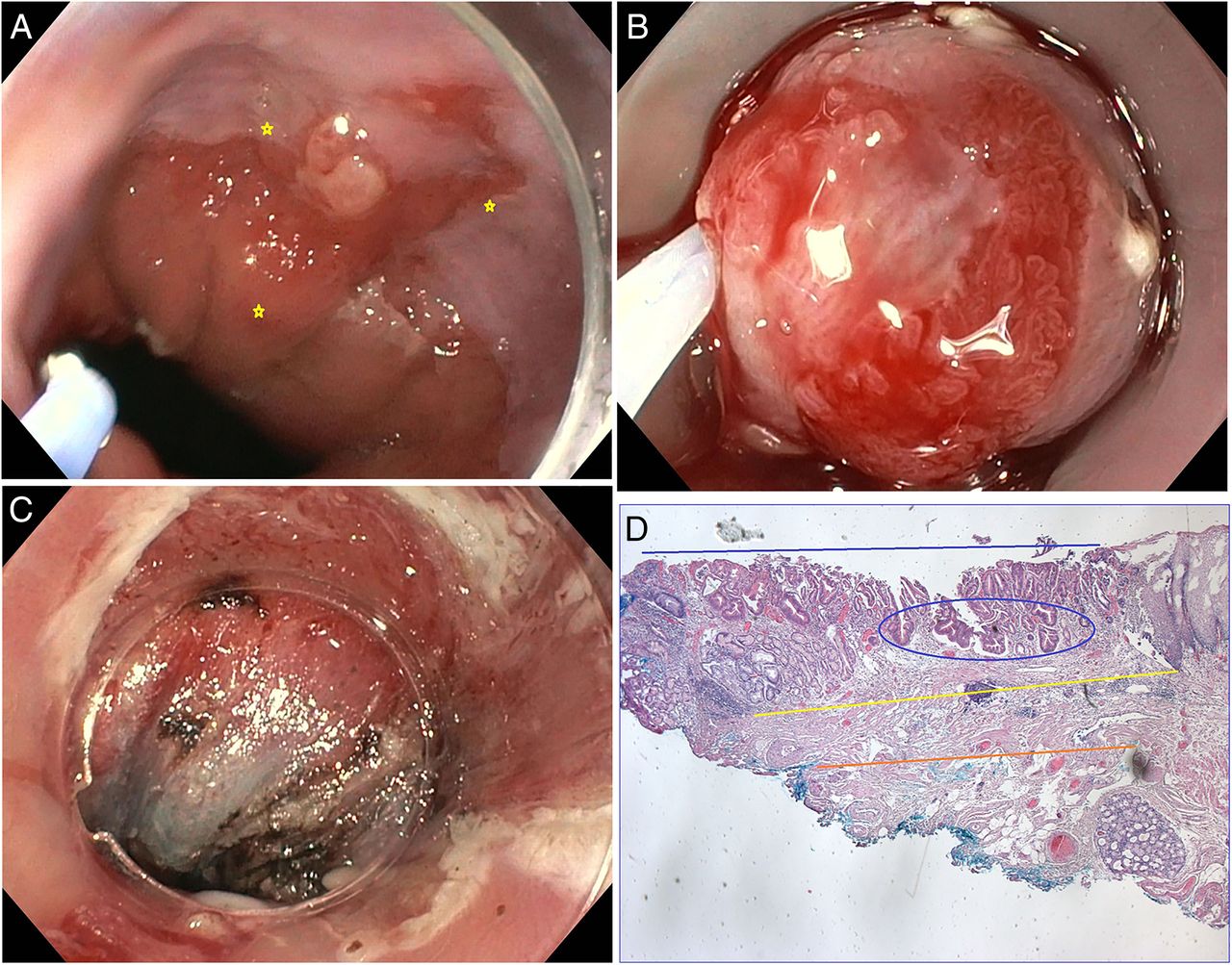
(A) Early Barrett adenocarcinoma, type 0–IIa and IIc (between yellow markers). (B) Endoscopic mucosal resection (EMR) showing part of the lesion including coagulation markers in the resection cap. (C) Area after complete resection of the lesion by piecemeal EMR. (D) Histology of one of the resected specimen showing mucosal adenocarcinoma pT1a (m1), L0, V0, tumour cell dissociation=0, pNX, R1 (HM1, VM0) G1 (blue bar: extension of AC, blue circle: deepest vertical tumour margin, yellow bar: upper muscularis mucosae, orange bar: lower muscularis mucosae).

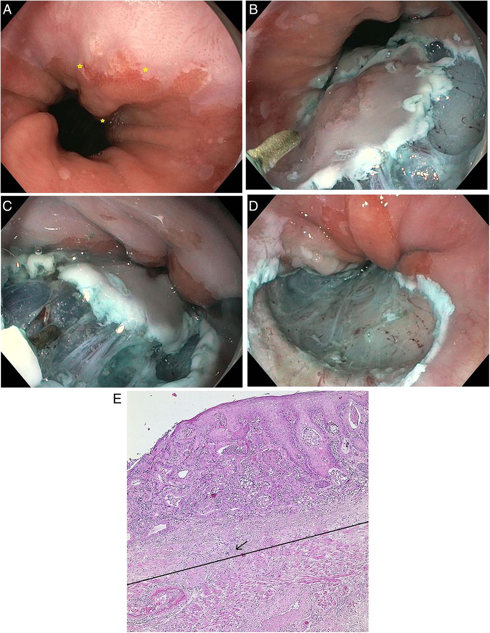
(A) Early Barrett adenocarcinoma, type 0–IIa and IIc (between yellow markers). (B) Endoscopic submucosal dissection (ESD) with circumferential incision of the mucosa at the periphery of coagulation markers with the HybridKnife. (C) Dissection of the submucosal layer by injection of saline solution with indigocarmine and subsequent cutting. (D) Area after complete en-bloc resection of the lesion by ESD. (E) Histology of the resected specimen showing mucosal adenocarcinoma pT1a (m2), L0, V0, tumour cell dissociation (TCD)=0, pNX, R0 (HM0, VM0) G2 (bar: upper layer of muscular mucosa; arrow: tumour cell complex invading the upper layer of the muscularis mucosae).
Intraprocedural bleeding was frequently observed but could be always stopped by endoscopic measures. ESD of deeper layers of the submucosa due to inappropriate lifting, suspicion of tumorous invasion or a technically difficult approach may explain transmural tears in three cases and perforation in two cases, respectively. Tears could be closed with hemoclips and patients remained asymptomatic. The two cases of perforation were registered as severe AE. In one of the patients, a perforation could be closed with three haemoclips after complete resection of the targeted lesion. Postoperative CT scan showed emphysema of the mediastinum and neck. There was no evidence of mediastinitis and the clinical course was uneventful under prophylactic antibiotic treatment. Hemoclips were also used in the other case with perforation and achieved closure of the defect according to endoscopic criteria. This patient developed retrosternal pain, fever and short breathing after 2 days. CT scan of the thorax showed extramural fluid collection at the site of resection and a right-sided pneumothorax. Endoscopy revealed a perforation at the distal margin of the resection area, which could be closed by application of an over-the-scope-clip (Ovesco Endoscopy AG, Tuebingen, Germany). Mediastinitis responded to additional treatment with parenteral nutrition, application of antibiotics and a thoracic tube for suction. The further clinical course was uneventful. Three additional patients in the ESD group and two in the EMR group had temporary chest discomfort. No further AEs were registered during hospitalisation or at 30 days according to telephone interviews. Routine endoscopy before discharge of the patients did not reveal any signs of perforation, acute bleeding or large vessels at the resection site, which required treatment. There was no 30-day or hospital mortality.
Histology of resected specimen and resection outcomes
Histology of the resected specimen revealed intestinal metaplasia in all cases but unexpectedly no neoplasia in four (table 3). Biopsy studies of the referring institution had shown adenocarcinoma in three of these cases and HGIN in one patient, which had been confirmed by an independent second histopathologist. Follow-up endoscopy did not indicate residual or recurrent neoplasia in these cases. Of the resected specimen, LGIN was diagnosed in two patients in the ESD group and HGIN in two cases in the EMR group.
Histology of resected specimen
There was no significant difference between both groups in the number of cases, grading and local staging of adenocarcinoma, which was diagnosed in all other cases. Submucosal invasion of adenocarcinoma was seen in 10 specimen with limitation to the superficial layer (sm1) in 8 of them. There was no case with infiltration of lymphatic vessels or veins.
Complete resection (R0) as the primary end point was confirmed in 10 of the 17 patients (58.8%) with HGIN or EAC in the ESD group. The rate was significantly higher compared with the EMR group (2/17; 11.8%) (p=0.01). The rates of curative resection including cases with sm1 infiltration but other low-risk factors fulfilled was 52.9% in the ESD group and 11.8% in the EMR group (p=0.03), respectively. Histological incomplete resection of HGIN or EAC was more frequently registered after EMR (n=15/17) than after ESD (n=7/17) (p=0.01). It was limited to the lateral tumour margin in 18 out of the 22 cases. Submucosal invasion of EAC was seen in all of the four cases with incomplete resection at the vertical margin.
Follow-up of >30 days
Elective oesophagectomy was performed in all of seven patients to whom surgery was recommended (table 4). There was no significant difference in the number of patients between both groups. Surgery was performed in four patients after ESD because of undifferentiated EAC (G3) with sm1 invasion (n=3) or deeper invasion of the submucosa (n=1). In the EMR group, three patients underwent oesophagectomy because of undifferentiated EAC (G3) with sm1 invasion (n=2) or deeper invasion of the submucosa (n=1). All of the four patients of the study in whom ESD or EMR could not achieve vertical tumour-free margins had at least one additional histological risk factor with indication for surgery. Histology of the oesophagectomy specimen showed no residual AC (n=4), residual mucosal AC (n=1) or a small area (diameter of 1 mm) of submucosal AC (n=1) with no lymph node metastases and no infiltration of lymphatics or veins in six out of the seven patients. One of these patients died at the age of 49 years 13 days after oesophagectomy due to severe adverse events (SAE). Histology had shown no more cancer after R0 resection by ESD of an undifferentiated EAC with sm1 invasion. Lymph node metastases were diagnosed in only one of the operated patients in whom histology of an ESD specimen revealed infiltration of vertical tumour margins due to massive submucosal invasion.
Follow-up of >30 days
All of the non-operated patients underwent at least one follow-up examination (table 4). Only one patient was subsequently lost to further follow-up after curative EMR of a mucosal EAC. Endoscopy had confirmed CR of neoplasia and intestinal metaplasia at 3 months. The 80-year-old patient with a health status ASA 3 did not wish further examinations in view of the excellent prognosis with regard to BO adenocarcinoma. All remaining patients (n=16 in each group) have been under surveillance according to the study protocol over mean period of 23.1±6.4 months.
CR of neoplasia was achieved after a single session of resection in 15 of 16 patients (93.8%) in the ESD group and 16 of 17 patients (94.1%) in the EMR group (p=1.0). Residual neoplasia was diagnosed in one patient in each group at the first follow-up examination. In the ESD group, histology of the resected specimen had shown well-differentiated mucosal EAC with incomplete resection at the lateral tumour margin. Endoscopy at 3 months showed a regular resection scar and no focal lesion. However, histology of biopsies of the scar area revealed HGIN. The remaining Barrett’s epithelium was subsequently resected by EMR with no histological evidence of neoplasia. In the patient of the EMR group, follow-up endoscopy revealed a small focal neoplastic area at the margin of a scar after histological incomplete resection of HGIN at the lateral margins. The patient underwent EMR, which achieved curative resection of a well-differentiated mucosal EAC.
RFA was recommended to all patients with remaining intestinal metaplasia. Two patients in the EMR group did not want to undergo additional interventions and preferred surveillance only. RFA was applied in all other cases and has so far achieved CR from intestinal metaplasia (IM) in 11 cases. The treatment is ongoing in four patients (table 4). No delayed AEs, in particular, no oesophageal stenosis, were observed in this study. Only a single case of a recurrent or metachronous neoplastic lesion was registered in this trial. The patient in the ESD group who had undergone successful retreatment of a histological residual HGIN at the resection scar developed a local recurrence of moderately differentiated EAC, which was detected 16 months after the second treatment. The lesion was completely resected by EMR and the first FU endoscopy at 3 months showed again local remission from neoplasia.
Discussion
EMR is currently considered to be the method of choice for resection of visible neoplastic lesions in BO because it is technically easy, safe and effective. The problem of recurrences can be overcome by RFA of the remaining BO epithelium. This combined approach was recently studied in 132 patients with early BO neoplasia in a European multicentre trial. CR from neoplasia and intestinal metaplasia was achieved per protocol in 98% and 93% of these cases, respectively. Neoplasia and IM recurred in 4% and 8% after a median of 27 months following the first-negative post-treatment endoscopic control.36
In spite of these excellent results of EMR of early BO neoplasia, ESD may offer advantages over EMR in terms of higher rates of R0 resection, a more detailed histological diagnosis and less recurrences. On the other hand, ESD is technically demanding and associated with more AEs.15 ESD of early neoplastic lesions in BO has not yet gained wide acceptance. Only two prospective and three retrospective uncontrolled single-centre trials have so far been published.17–21 Details in table 5 show various results probably due to different selection criteria, procedural techniques and definition of outcome parameters. Therefore, the discussion of advantages and disadvantages of EMR or ESD of early BO neoplasia is controversial and a randomised trial seemed to be necessary.22 ,23
Results of published trials on endoscopic submucosal dissection for early Barrett's oesophagus neoplasia
The present study represents the first randomised comparison between EMR and ESD for endoscopic treatment of early BO-related neoplasia. We are aware of several limitations of this study, which is an open trial where examiners cannot be blinded to the technique used and may favour one technique or the other. We, however, tried to minimise this bias by randomising patients after inclusion and marking of the lesions so that biases excluding non-favourable lesions for one specific technique can be excluded. We also determined the technical steps for each procedure in detail to minimise bias. Classifications used for endoscopic and histopathological classification of resected lesions may have some inter-rater variability, but we tried to minimise this information bias since randomisation was done after classification and pathologists were blinded to the resection technique in terms of the resected specimen and biopsies of follow-up studies. R0 resection was determined as the primary outcome parameter. According to published series on EMR and ESD, R0 is more frequently achieved by ESD. It should facilitate the histological evaluation of the specimen and could increase rates of remission from neoplasia and decrease the risk of recurrences compared to R1 resection. We alternatively considered clinically oriented parameters, for example, CR of neoplasia or recurrences as primary objectives. CR from neoplasia was observed on average in 95% of the cases after EMR as well as after ESD.3–14 ,17–21 We realise that the outcome of recurrence or recurrence-free survival would have been preferable, but would require very large patient numbers. Even under consideration of a favourable outcome of ESD with CR of neoplasia in 98% compared with 92% as one of the lower reported rates after EMR, 523 patients would have to be included in a randomised trial to show a significant difference between both methods at a level of 5%, a power of 80% and an overhead of 10%.6 ,19 This is supported by our results, where recurrence rates were very low (one case in each group). So, if this study would be taken as basis of a power calculation of a possible outcome study, case numbers would be even much higher. However, we acknowledge the choice of a surrogate outcome parameter as a further limitation of this randomised trial.
Recurrences are probably less frequent in patients who underwent en-bloc resection by ESD. However, it can be difficult to differentiate between a recurrent or a metachronous neoplastic lesion and details were usually not reported. In addition, it seems no longer justified to follow patients after ER of neoplasia without eradication of remaining IM.1 Given the low recurrent neoplasia of 4% after EMR plus RFA, it is unlikely that replacement by ESD will lead to a significant reduction: recent trials have shown recurrence rates of 2–10% after ESD even in combination with additional interventions in case of residual IM18–21 ,36 (table 5).
Most of the inclusion and exclusion criteria of the present study are comparable to other trials on ER. In contrast to other series, we excluded patients with visible flat lesions because of the very low risk of adenocarcinoma with submucosal invasion and the difficulties to delineate the lateral tumour margin, which is mandatory to achieve RO resection. We also excluded patients with multifocal neoplastic lesions or lesions >3 cm in the longitudinal direction or extending to more than half of the oesophageal circumference. Several resections or widespread resections by piecemeal EMR or ESD have to be performed in these cases, and the comparison would again require inclusion of a large number of cases.
The characteristics of the 40 included patients and the neoplastic lesions were not significantly different between both groups (table 1). The mean diameter of the tumours was similar to other series on ESD (table 5). In contrast to other trials, the majority (92%) of the procedures were performed under deep sedation without endotracheal intubation. According to our protocol, patients were randomised after marking of the lateral tumour margin so that routine general anaesthesia would have been applied in all patients. This approach was not considered justified for EMR, which can be safely performed under sedation. In case of ESD, a precise control of the knife became sometimes difficult under sedation in particular at the gastro-oesophageal junction when patients breathed deeply and irregularly or if they were restless. This limitation had no impact on the completeness of resection but could have increased the risk of perforation. The mean procedural duration was significantly longer for ESD compared with EMR (p=0.0002) (table 2). However, ESD took less than half the time compared with most of the other trials. This difference may be explained by smaller areas of resection and by the water-jet-assisted technique of ESD. We could recently demonstrate that water-jet-assisted ESD is significantly faster than conventional ESD for early gastric cancer and requires less frequently exchanges of accessories.28 ESD as well as EMR achieved endoscopically complete resection of the targeted areas in all cases. However, en-bloc resection could be always obtained by ESD but in only 3 of the 20 cases by EMR (p<0.0001). Rates of en-bloc resection were rarely reported for EMR in other trials probably because piecemeal resection is usually required for complete removal of even smaller neoplastic lesions including all coagulation markers. The maximal diameter of the largest specimen (mean±SD: 29±9 mm) was higher in the ESD group compared with EMR (mean±SD: 18±4 mm; p<0.0001). However, it was smaller than in other trials on ESD for early BO neoplasia although the size of the targeted areas was similar (table 5). This difference implies larger safety margins for resection that were not reported or varied between 3 and 10 mm in recent trials compared with 3 mm in the present study.
There was no significant difference in rates of AEs between both groups. Severe AEs were only registered in the ESD group. They were related to oesophageal perforation in two patients. They could be closed with hemoclips in both cases but caused mediastinitis in one patient who reassumed eating solid food after 2 days. The perforations were caused by inadvertent cutting in the muscle layer close to the cardia, which may have been caused by unexpected deep breath or restlessness of the patients under sedation. Perforation was less frequently observed when ESD was performed under general anaesthesia in other trials (table 5).
No delayed AE was observed in the present trial. There was no stenosis that caused symptoms or required interventions. In contrast, the incidence of stenoses after ESD of early BO neoplasia ranged from 9% to 60% in studies from other centres, which are most probably caused by wider resection (table 5).19 ,21 On average, four sessions of dilations were required for the management of stenoses in the study with the highest incidence.21 These results suggest that stenoses are not caused by the technique of ESD but they are due to wide resection as it was shown for widespread EMR.34
Histology of the resected specimen showed no dysplasia or low-grade dysplasia in three patients in each group. HGIN or adenocarcinoma had been diagnosed from biopsy specimen by two independent histopathologist before study entry. This discrepancy could be explained by small neoplastic areas in focal lesions, which had been removed with biopsies or by false positive findings due to inflammatory changes. Alternatively, the neoplastic area may have not been resected, which is unlikely because no residual neoplasia was diagnosed at the follow-up examinations. In another trial, histology of specimen resected by widespread ESD did not confirm HGIN or EAC in 10% of the patients.21 Others excluded these cases from the study after resection.19 In the present study, HGIN or EAC was diagnosed in 17 patients in each group. Infiltration of the submucosa was seen in one-third of the 32 patients with adenocarcinoma. Lower rates varying between 9% and 16% were reported in other series.19–21 In contrast to these trials, we had excluded patients with flat neoplastic lesions because of the very low risk of advanced histology. The potential advantage of ESD over EMR is less obvious in these cases, which even meet criteria for primary RFA.1 ,2
The primary objective of the study, R0 resection of the targeted neoplastic area, was significantly more frequently achieved by ESD compared with EMR (58.8% vs 11.8%; p=0.01), which can be explained by the frequent need of piecemeal resection for EMR that does usually not allow to confirm lateral tumour-free margins. These rates of R0 resection were lower for both groups than the rates that were adopted for the calculation of the sample size (80% for ESD and 30% for EMR, respectively). The statistical power for the actual resection rates, 20 patients per group and a level of significance of 5% lead to a statistical power of 82.9%. Therefore, the study is not underpowered and the statistical statements are valid.
R1 resection at the basal margins was only diagnosed in four patients of the study in whom EAC infiltrated the submucosa. Higher rates of R0 resection (64–85%) of ESD of HGIN or EAC were reported by other authors18–21 (table 5) in spite of comparable en-bloc resection rates. Therefore, the better results could be best explained by a more precise delineation of tumour margins or more likely by larger safety margins for resection. However, although the mean diameter of the specimen was 53 mm for a mean targeted area of 20 mm, the R0 resection rate was not >64% in one study.21 This moderate advantage of wider resection has to be balanced against doubling of the procedural time and the development of oesophageal strictures in 60% of the cases compared with none in our trial. The curative resection rates were also higher in the ESD group compared with the EMR group (52.9% vs 11.8%; p=0.03) mainly due to more cases with horizontal R0 resection. These results indicate that the term ‘curative resection’, although reported in the majority of trials on ESD, is not useful from a clinical standpoint. In particular, all cases with lateral positive margins are considered as ‘non-curative’ although follow-up studies and data of the current trial indicate complete local remission of neoplasia in the majority of these cases.
The parameters that indicated the need for elective surgery according to our study protocol, for example, grading, determination of lymphatic or venous vessel infiltration and the depth of vertical tumour invasion, could be well determined in both groups. There was no significant difference in the number of patients who underwent oesophagectomy after ESD (n=4) or EMR (n=3) (p=1.0). The rate of indication for surgery after ER of early BO neoplasia was 21%, which is comparable to rates between 9% and 36% in recent trials on ESD for the same indication.18–21 These data cannot be compared with studies on EMR because of different inclusion criteria. However, the present study indicates that it seems to be very unlikely that ESD is more accurate than EMR for the identification of patients who benefit from surgery. The very low risk of missing more advanced tumour stages after EMR of EAC can be also concluded from the long-term follow-up of a previous study, which identified only 2 out of 1000 patients who died from BO adenocarcinoma after EMR.14
We have to say at this point that our main outcome was merely technical and en-bloc R0 resection may not be an ideal oncological surrogate marker. This was shown by our study since CR from neoplasia of the non-operated patients was seen in all but two of our patients after a single session of ESD or EMR, respectively. Both patients with small residual lesions after R1 resection were successfully treated by EMR. These excellent results correspond to other studies on ESD and also EMR. However, rates of >90% of complete local remission from neoplasia required more than one session in several previous trials on EMR.3 ,7 ,8 CR from intestinal metaplasia was observed in 16 of 33 patients (48.5%) with no significant difference between ESD or EMR (table 4). Similar rates were reported in other trials in spite of wider resection by ESD (table 5). These results suggest that approximately every second patient has to undergo additional interventions from complete eradication from IM independently from the resection technique to reduce the risk of recurrent or metachronous neoplasia.35 ,36 This combined approach has been also used in other trials on ESD.17 ,19 ,21 The majority of the patients of the present trial with residual IM accepted to be treated by RFA. The treatment achieved eradication of IM in 11 of 15 cases and is still ongoing in four patients. Following this approach, there was only one patient in whom a local recurrence was diagnosed after re-treatment of a residual neoplasia during a mean follow-up period of 23.1±6.4 months. This low rate cannot be compared with previous trials on EMR, which did not consider additional treatment for residual non-neoplastic IM. However, the rate of CR from neoplasia of 97% in the non-operated patients does correspond to the result (98%) of a recent large multicentre trial of a combined treatment of early BO neoplasia by EMR and RFA.36 The high rates in both groups of the present trial indicate that a large number of patients would have to be randomised to show a potential significant difference between EMR and ESD for this outcome parameter.
In conclusion, ESD and EMR are both highly effective for complete ER of early BO neoplasia. Compared with EMR, ESD is more time-consuming and the risk of severe AE may be higher. On the other hand, ESD more frequently achieves en-bloc, R0 and curative resection. However, corresponding to results of uncontrolled trials this advantage does not seem to have an impact on the need for elective surgery or rates of CR from neoplasia. The study cannot exclude that en-bloc resection by ESD can reduce the risk of missing important histological details in selected cases of more advanced tumour stages compared with piecemeal EMR. Recurrent or metachronous neoplasia is rare after ESD as well as EMR provided that RFA is performed for eradication of residual non-neoplastic Barrett’s epithelium. The risk of stricture formation after ESD seems to be low if the resection area is limited to focal lesions and <50% of the oesophageal circumference. In view of the similar excellent outcomes after ESD and EMR of early BO neoplasia in this trial and recent uncontrolled series, it seems very unlikely that even large-scale trials will show significant differences in clinical long-term outcome parameters between both methods.
Acknowledgments
The authors thank Erbe Elektromedizin GmbH, Tuebingen, for providing the HybridKnife instruments without charge.
References
Footnotes
Correction notice This article has been corrected since it published Oline First. The OA licence has now been added.
Contributors GT: conception and design, acquisition and interpretation of data, critical revision of manuscript. EMH: enrolment of participants, assignment of participants acquisition, analysis and interpretation of data, critical revision of manuscript. MV, HG: study conduction, interpretation of data and critical revision of manuscript. ME: conception and design and critical revision of manuscript. AN: generation of the random allocation sequence, data analysis and critical revision of manuscript. BS: conception and design, study conduction, critical revision of manuscript. HN: conception and design, study conduction, writing of the first draft of the manuscript.
Competing interests None declared.
Patient consent Obtained.
Ethics approval The study protocol was approved by the International Medical and Dental Ethics Commission GmbH in Freiburg, Germany (an institution registered at the Office of Human Research Protections of the US Department of Health and Human Services and at the German Federal Institute for Drugs and Medical Devices). The study was performed in compliance with the Declaration of Helsinki and good clinical practice.
Provenance and peer review Not commissioned; externally peer reviewed.