Article

A Pooled Analysis of Serum Phosphate Measurements and Potential Hypophosphataemia Events in 45 Interventional Trials with Ferric Carboxymaltose

1
Department of Medical Sciences, IRCCS San Raffaele Pisana, 00166 Rome, Italy
2
Department of Gastroenterology, Hepatology, Diabetology and Endocrinology, Klinikum St. Georg, 04129 Leipzig, Germany
3
Clinical Development, Vifor Pharma, 8152 Glattbrugg, Switzerland
4
Biometrics, Vifor Pharma, 8152 Glattbrugg, Switzerland
5
Department of Human Sciences and Quality of Life Promotion, San Raffaele University, 00166 Rome, Italy
6
Unit of Clinical and Molecular Epidemiology, IRCCS San Raffaele Pisana, 00166 Rome, Italy
7
Department of Gastroenterology and Clinical Nutrition, DGD Clinics Sachsenhausen, 60594 Frankfurt am Main, Germany
*
Author to whom correspondence should be addressed.
J. Clin. Med. 2020, 9(11), 3587; https://doi.org/10.3390/jcm9113587
Submission received: 29 September 2020 / Revised: 2 November 2020 / Accepted: 4 November 2020 / Published: 6 November 2020
(This article belongs to the Section Hematology)

Abstract

:
Ferric carboxymaltose (FCM) has been shown to achieve rapid replenishment of iron stores and correction of anaemia in various populations with iron deficiency. A decrease in serum phosphate (PO43−) levels, which in most cases is asymptomatic, has been reported with IV iron preparations. Hypophosphataemia (HP) is a known adverse drug reaction with FCM. This post hoc pooled analysis investigates the frequency, duration, risk factors, and clinical signs of HP as reported in interventional clinical trials with FCM. Pooled data from subjects enrolled across 45 clinical trials in different therapy areas were included. A three-step adjudication process was utilised to identify adverse events of HP. Stratified analyses by therapy group and stepwise logistic regression analysis were used to identify predictors of HP. This pooled analysis confirms that FCM is associated with increased rates of serum PO43− lowering, but mean serum PO43− values were seen to recover at Week 4 and further recover at Week 8. Among all subjects receiving FCM therapy (n = 6879), 41.4% (n = 2847) reached a PO43− nadir value <2.5 mg/dL at any point on study and 0.7% (n = 49) reached a nadir <1 mg/dL. Although gastroenterology and women’s health subjects were identified to be at higher risk, occurrence of severe HP (<1 mg/dL [0.3 mmol/L]) following FCM administration was not observed to be common among subjects in these studies. Furthermore, there was no correlation between laboratory serum PO43− values and the occurrence of reported adverse events related to low PO43− levels.

1. Introduction

Hypophosphataemia (HP) is uncommon in the general population and is often an incidental finding, detected in 2–3% of hospitalised patients [1]. The normal range for serum phosphate (PO43−) is 2.5–4.5 mg/dL in adults (0.81–1.45 mmol/L). Mild, moderate, and severe HP are defined as a serum PO43− level of 2.0–<2.5 mg/dL (0.65–<0.81 mmol/L), 1–<2.0 mg/dL (0.32–<0.65 mmol/L), and <1 mg/dL (<0.32 mmol/L). However, extracellular fluid PO43− levels represent <1% of the total body phosphorus content and are not a good index for PO43− stores in bones or soft tissue [2,3,4]. Mild HP is usually asymptomatic. Mild or moderate HP of short duration generally does not require treatment. Severe cases with PO43− depletion may cause symptoms such as fatigue [5], myocardial depression [6], rhabdomyolysis [7], seizures [8], and haemolytic anaemia [9] and, over time, can ultimately contribute to bone abnormalities such as osteomalacia or rickets [10,11]. Rarely, the most severe cases of HP can be fatal [12,13,14]; respiratory muscle dysfunction or impaired myocardial metabolism and decreased cardiac contractility have been associated with severe HP [6,13,15,16].
Hypophosphataemia is a known adverse drug reaction with ferric carboxymaltose (FCM) listed in the Ferinject® (FCM) Prescribing Information since registration of the product [17], and it is often transient and, in most cases, asymptomatic. A decrease in serum PO43− has also been reported with several other IV iron preparations [18,19,20]. However, the potential clinical relevance of a decrease in serum PO43− has been challenging to establish due to the generalised symptoms of HP. The mechanisms of FCM-induced HP are still to be fully elucidated, although it is probably attributed to the intracellular metabolism of fibroblast growth factor 23 (FGF23). Initially attributed to regulating PO43− and calcium homeostasis, recent studies have also linked FGF23 to iron homeostasis [21,22,23,24] potentially leading to HP via increased PO43− loss in the urine [21]. Although replenishing iron stores appears to reduce the production of FGF23, IV iron preparations such as FCM may potentially inhibit the proteolytic cleavage of FGF23, thereby increasing the circulating levels of iFGF23 [25], which in a downward cascade, may have an impact on phosphate homeostasis causing phosphaturia. The exact mechanisms mediating iFGF23 increase by FCM is unknown [26].
FCM has been studied extensively in both the clinical trial and real-world settings (>12 million patient-years of post-marketing exposure) [27] and is an effective and generally well-tolerated treatment to rapidly replenish iron stores and correct anaemia in patients with iron deficiency of various aetiologies [28]. FCM is an intravenous iron preparation in which a non-dextran, stable carbohydrate shell facilitates iron release in a controlled manner. As a result, a single high-dose course of FCM (up to 2 × 750 mg of iron in the USA and 1 × 1000 mg of iron in Europe) can be administered over 15 min, allowing rapid iron repletion even in severely iron-deficient patients.
FCM has been shown to achieve rapid replenishment of iron stores and correction of anaemia in various populations with iron deficiency and to be more effective than oral iron therapy or iron sucrose [29,30]. It is generally well tolerated, with a lower risk of severe hypersensitivity reactions compared with some other IV iron formulations [29,31,32,33].
The purpose of this post hoc pooled analysis is to investigate the incidence and extent of HP in FCM-treated patients and to characterise the frequency, duration, risk factors, and potential clinical signs of HP as reported in interventional clinical trials.

2. Methods

Analyses were based on the pooled study populations of all clinical trials with FCM that had individual patient data filed in the clinical databases of Vifor Pharma and licensing partners (data lock point: 31 October 2017) [27]. Study duration varied from 1 to 52 weeks. Observational studies and studies without a control group were excluded. For studies with a cross-over design, only data from the first treatment period were analysed. The list of all studies contributing data to the pooled analysis is reported in Supplementary Table S1. Serum PO43− levels were defined according to Common Terminology Criteria for Adverse Events (CTCAE) thresholds (v4.0) and severity definitions (v5.0) as follows: 2.5–<4.5 mg/dL (normal), 2.0–<2.5 mg/dL (mild decrease), 1–<2.0 mg/dL (moderate), and <1 mg/dL (severe) [34,35].
All individual subjects from the selected studies were pooled and analysed. In total, 15,080 subjects (FCM, n = 8245 [54.7%], controls, n = 6835 [45.3%]) from 45 clinical trials were included in the safety analysis (safety population). Control groups included subjects who received the following treatments: other IV iron (n = 1998), oral iron (n = 1621), placebo (n = 616), and standard medical care (n = 2600). All analyses evaluating the modification of serum PO43− levels throughout study duration included the 6879 subjects with post-baseline serum PO43− measurements (83.4% of all subjects receiving FCM).
Stratified analyses were performed by nephrology, cardiology, gastroenterology, neurology, women’s health, and other therapeutic areas. Variables considered to be potential confounders or effect modifiers of the possible relationship between FCM and HP included: gender; age (as a continuous variable categorised as <18, 18–65, ≥65 years); baseline BMI (as a continuous variable categorised as normal, overweight, obese); baseline anaemia (yes/no); baseline ferritin (µg/L); and baseline transferrin saturation (TSAT) (%). Effects of FCM treatment were also considered: single vs. multiple dose (n [%]); maximum single dose (categorised as ≤15 mg/kg and >15 mg/kg); and cumulative dose (n [%]) (categorised as ≤1000 mg, >1000–≤1500, >1500 mg).
Adjudication of HP adverse events (AEs): investigator-reported AEs containing any of the following MedDRA Preferred Terms (version 20.1) were positively adjudicated as HP: blood phosphorus decreased, hypophosphataemia, hypophosphataemic rickets, and hereditary hypophosphataemic rickets. A set of 318 MedDRA Preferred Terms (including, but not limited to, fatigue, muscle weakness, muscle pain, bone pain, osteomalacia, haemolysis, white cell dysfunction, neurological symptoms, cardiac failure, and ventricular tachyarrhythmia) were interrogated and cross-checked against time to onset, possible treatment, and laboratory PO43− values to adjudicate AEs as HP related to FCM. The algorithm for adjudication of an AE of HP is described in Supplementary Figure S1.
Statistical analysis: All statistical analyses were performed using SAS version 9.4 (SAS Institute Inc., Cary, NC, USA). Mean values of baseline PO43− and corresponding 95% confidence intervals (CIs) were used to determine the extent and timings of change in PO43− from baseline values by plotting residual, leverage, and influence measures as diagnostic quantities.
Univariate logistic regression was first performed to select potential risk factors (p ≤ 0.20) then the set of covariates predicting the risk of HP was estimated with a multiple logistic regression model, providing odds ratios and the corresponding CIs for the risk of developing HP associated with FCM treatment.

3. Results

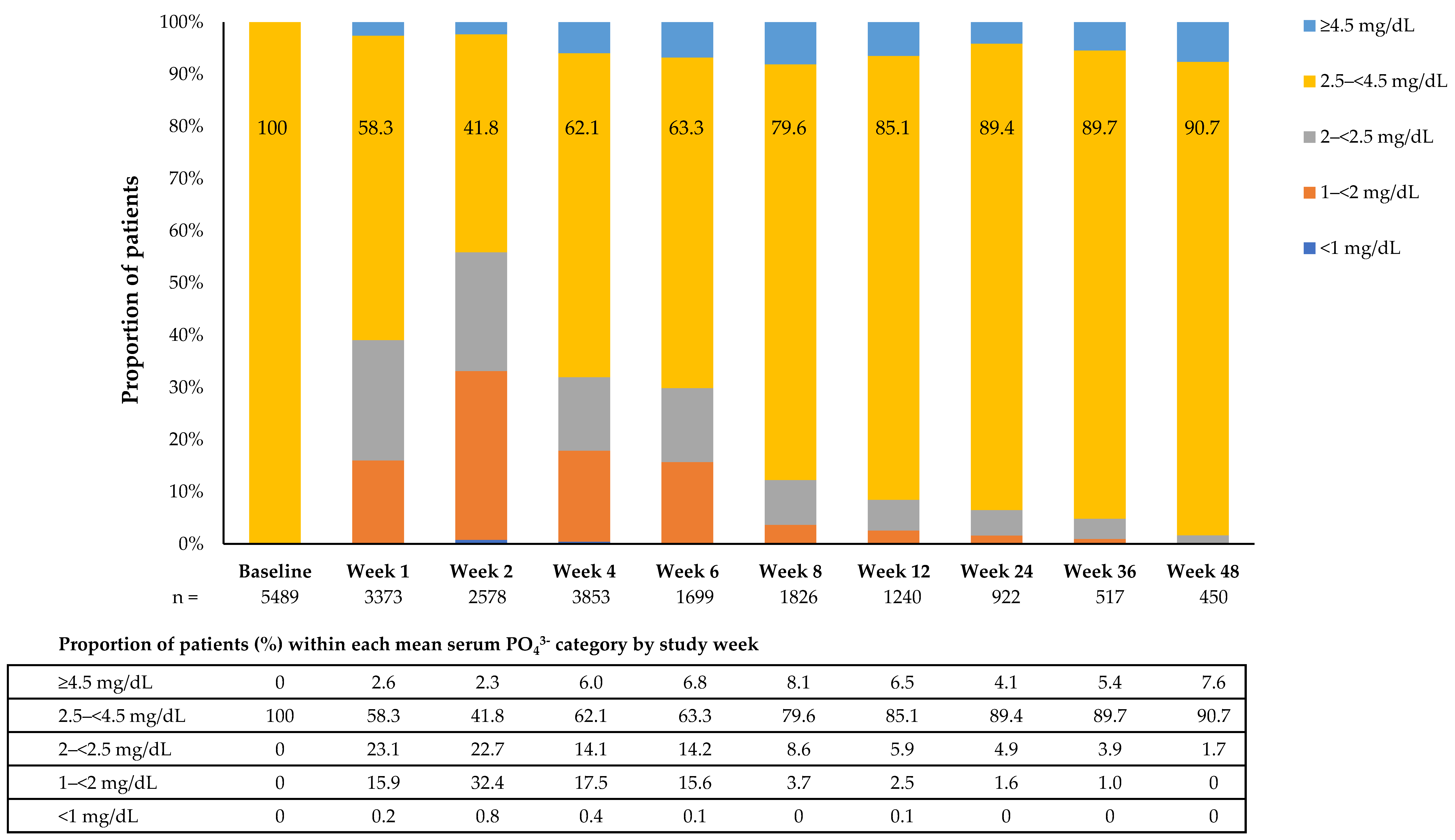
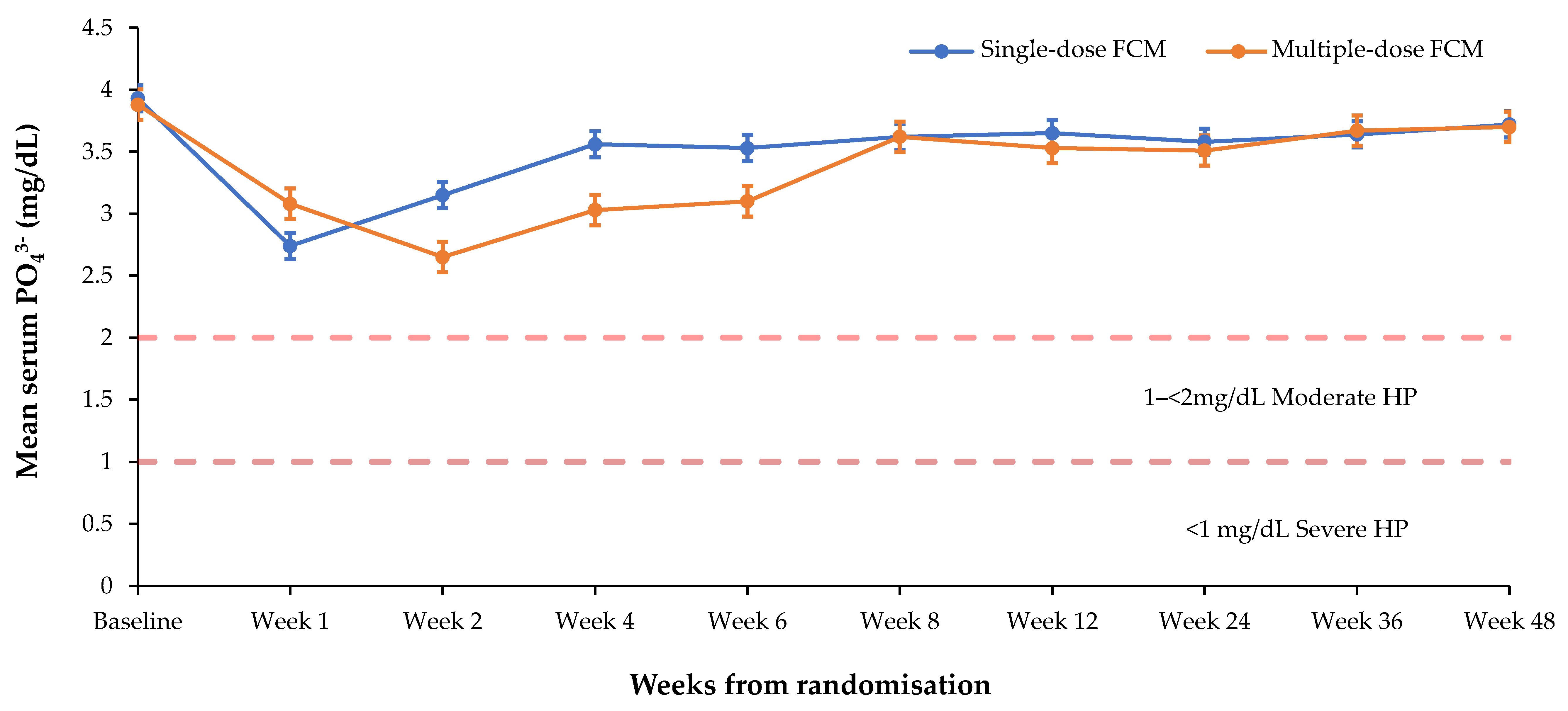
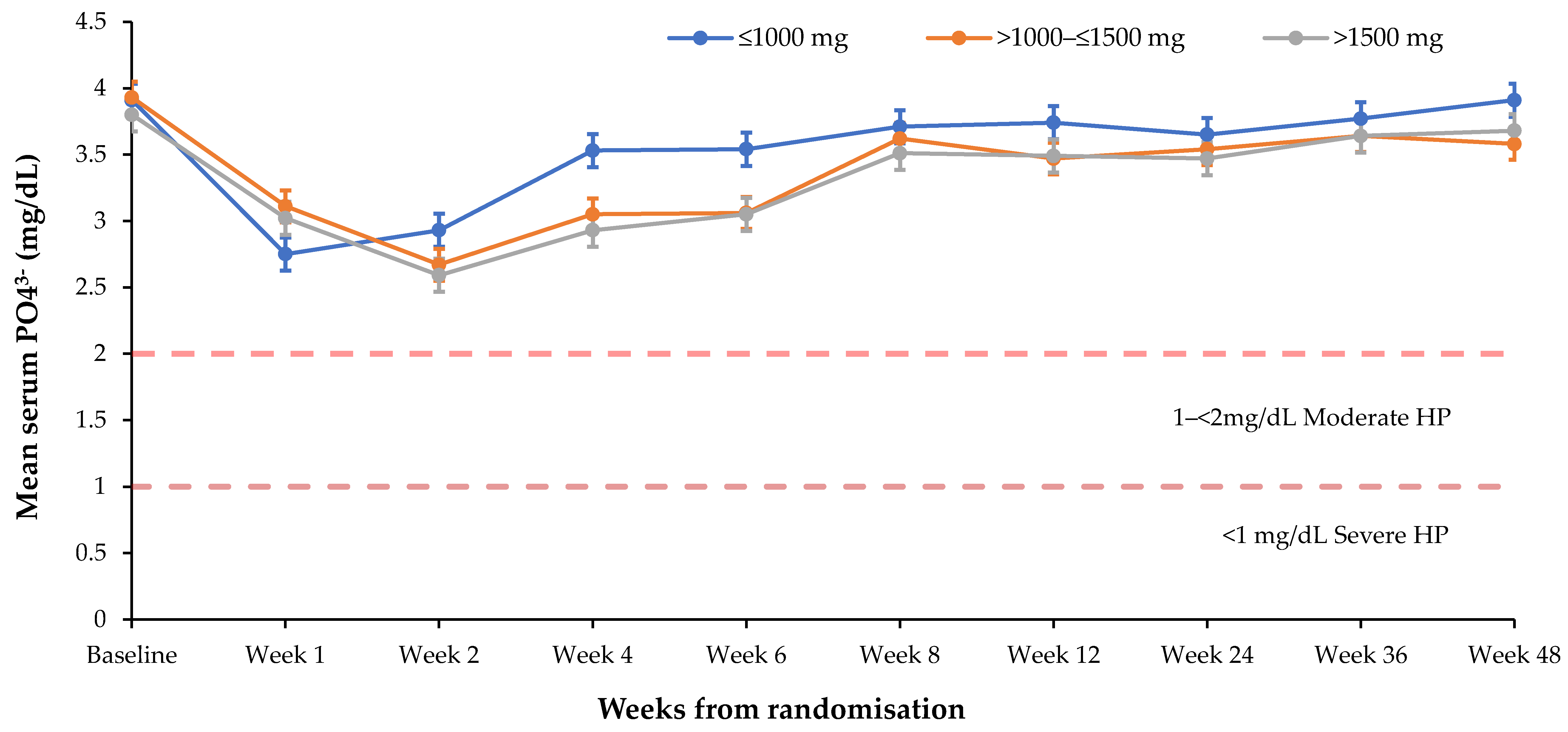
3.1. Effect of FCM on Serum PO43− Levels

At baseline, 90.4% of patients receiving FCM therapy (n = 6879) were anaemic, mean serum ferritin was 43.5 µg/L, mean TSAT was 14.3%, and 98.2% of patients had serum PO43− values ≥2.5 mg/dL (Supplementary Table S2). Baseline mean serum PO43− varied across therapy areas; the proportion of patients with mean PO43− < 2.5 mg/dL ranged from 2.0% (gastroenterology) to 0.6% (non-dialysis-dependent chronic kidney disease (NDD-CKD)). Baseline mean ferritin ranged from 20.2 µg/L (women’s health) and 21.2 µg/L (gastroenterology) to 77.6 µg/L (NDD-CKD) and 109.9 µg/L (haemodialysis-dependent chronic kidney disease (HD-CKD)).
Reduction in mean serum PO43− was observed from the first week of treatment with FCM (n = 6848), reaching a nadir at Week 2 (Figure 1). This trend was observed to a lesser extent in the other IV iron group but not observed in any of the other control groups (Oral iron, Placebo, Standard Medical Care (Table 1)).
Mean change from baseline in serum PO43− was greatest at 2 weeks; mean baseline PO43− of 3.9 (SD 0.88) mg/dL decreased by 1.24 (SD 0.93) mg/dL, which corresponded to a mean PO43− level of 2.69 (SD 1.08) mg/dL. However, by Week 4 mean serum PO43− levels had recovered to 3.22 mg/dL and recovered further at Week 8 to 3.62 mg/dL.
Among all subjects receiving FCM therapy (n = 6879), 41.4% (n = 2847) reached a PO43− nadir value <2.5 mg/dL at any point during the study and 0.7% (n = 49) reached a nadir <1 mg/dL. Of the FCM subjects, 2599 (37.8%) with baseline PO43− 2.5–<4.5 mg/dL developed HP (PO43− <2.5 mg/dL) during the study, with 22.8% of patients reaching values <2 g/dL and 0.7% of patients <1 g/dL at any time point during the studies. Among subjects with normal PO43− values at baseline, 87.7% (1602/1826) of evaluable FCM patients at Week 8 had mean serum PO43− values ≥2.5 mg/dL (Figure 2). This trend was also observed in the total population, with 89.0% of evaluable FCM-treated subjects at Week 8 having mean serum PO43− levels ≥2.5 mg/dL (Table 2). These observations support the hypothesis that FCM treatment induces a temporary decrease in mean serum PO43− levels.

3.2. Risk Factors for Hypophosphataemia

The proportion of patients with moderate or severe HP was higher in the gastroenterology and neurology therapy areas (47.1% and 39%, respectively) and considerably less in cardiology, NDD-CKD, and HD-CKD patients (9.9%, 12.3%, and 0%, respectively) than in the total population. Among the 49 patients who reached a PO43− nadir below 1 mg/dL, more than half (55.1%) belonged to gastroenterology studies and 11 (22.4%) to women’s health studies.
Serum PO43− levels were lowered to a greater extent and remained low for a longer duration in patients receiving multiple vs. single doses of FCM (Figure 3) (nadir of 2.65 vs. 2.74 mg/dL), in patients receiving frequent (>1 administration during the first 4 weeks of treatment) vs. non-frequent dosing (nadir of 2.61 vs. 3.30 mg/dL), and in patients receiving higher cumulative FCM doses (Figure 4) (nadir of 2.59 mg/dL for >1500 mg vs. 2.67 for 1000–≤1500 mg and 2.75 for ≤1000 mg, respectively. The mean cumulative dose received was 1315 mg (n = 6879; min, max: 12.5 mg, 6500 mg).
Risk factors for developing post-baseline moderate HP and post-baseline severe HP within 12 weeks of treatment, identified by logistic regression analyses, included treatment setting, baseline ferritin, and FCM dosing scheme (Table 3). Among the factors tested, the most dominant risk factors for moderate HP only (i.e., subjects who did not progress to severe HP) were therapeutic area (neurology, gastroenterology, and others vs. women’s health), baseline ferritin level, baseline BMI, FCM cumulative dose, FCM single vs. multiple dose, and belonging to Black or African American race. The most dominant risk factors for severe HP were therapeutic area, particularly gastroenterology vs. women’s health (odds ratio (OR): 4.601 [2.093–10.111]), and FCM cumulative dose >1000–≤1500 mg vs. ≤1000 mg (OR: 5.867 [2.514–13.694]). Of note, maximum single dose was not associated with an increased risk of developing severe HP (p = 0.522).

3.3. Adverse Events Possibly Associated with Hypophosphataemia

3.3.1. Investigator-Reported Adverse Events of HP

Treatment-emergent AEs of HP occurred in 2.2% of subjects receiving FCM (n = 179, all non-serious) vs. 0.2% with other IV iron (n = 3), 0% with oral iron or placebo, and <0.1% with standard medical care (n = 2). Phosphate supplementation was administered to 35 of 179 subjects who had a reported AE of “hypophosphataemia” and “blood phosphorus decreased”. Of note, among 39 subjects with a low laboratory PO43− value at Week 12, one patient required phosphate supplementation.

3.3.2. Clinical Signs Possibly Related to HP

Cross-checking reported AEs against a defined pool of 318 MedDRA terms describing AEs possibly related to HP identified 8.8% (n = 726) FCM-treated subjects, 12.5% other IV iron (n = 249), 8.6% oral iron (n = 140), 14.0% placebo (n = 86), and 4.2% standard medical care (n = 109) (Table 4). Among all patients with a potential clinical sign of HP, two had a serious treatment-related AE, one (0.01% of the FCM safety population) in the FCM group (congestive heart failure, “other” study group) and one (0.16% of the placebo safety population) in the placebo group (chronic heart failure, “cardiology” study group).
Serious cardiac disorders classified as potentially related to HP occurred in 1.0% FCM (n = 81), 1.9% other IV iron (n = 37), and 0.6% oral iron subjects (n = 10) (Supplementary Table S3). Musculoskeletal and connective tissue disorders occurred in one FCM subject from the NDD-CKD nephrology treatment group (osteoporotic fracture). This occurred in an 80-year-old female and was not positively adjudicated as HP.
Four of 49 FCM subjects (8.2%; 0% serious) who had nadir PO43− below 1 mg/dL had a potential clinical sign of HP (increased C-reactive protein [CRP], fatigue, hypocalcaemia, elevated white blood cell value, swollen finger, tingling in hand) vs. 8.0% (126/1569) with 1–<2.0 mg/dL (0.5% serious), 9.2% (113/1229) with 2.0–<2.5 mg/dL (1.5% serious), and 10.2% (411/4032) with ≥2.5 mg/dL (2.3% serious). These data demonstrate no apparent correlation between laboratory PO43− values and the manifestation of AEs possibly related to HP.

3.3.3. Adjudicated Symptoms among Subjects with Investigator-Reported HP

Adjudicated HP AEs according to treatment type occurred in 0.4% (32) FCM vs. 0.5% (9) other IV iron, 0.4% (6) oral iron, 0% placebo, and <0.1% (1) standard medical care. None of these cases were serious. Within the FCM-treated group, four patients with an adjudicated AE of HP also had serum PO43− levels <1 mg/dL while on-study (two patients within the gastroenterology group and two patients from the women’s health therapy area).

4. Discussion

Following FCM administration, HP is a common occurrence [17]; severe HP (<1 mg/dL) and associated severe AEs are not common. The occurrence of severe HP following FCM administration was not observed to be common among subjects in the 45 analysed studies comprising 8245 subjects treated with FCM. Furthermore, there was no correlation between reported laboratory serum PO43− values and the reported occurrence of AEs.
The effect of serum PO43− lowering following administration of FCM has been frequently observed in interventional, observational, and retrospective studies [20,36,37,38,39,40,41,42]. This effect has been seen at 2 weeks following FCM administration, and persistence of laboratory HP at Weeks 5 and 6 has been noted [39,40,41]. Although a greater reduction in serum PO43− levels was observed with FCM vs. comparators in these studies, the shorter study duration of such reports precludes a fuller observation on the duration of this reduction in serum phosphate. The present pooled analysis of clinical studies confirms that FCM is associated with increased rates of serum PO43− lowering, but mean serum PO43− values were seen to recover at Week 4 and further recover at Week 8. Of the 1532 subjects with low serum PO43− levels at Week 2, only 39 such cases remained at Week 12 after the first dose, all of which were limited to mild or moderate levels. Intervention with phosphate supplementation was required for one of these 39 subjects.
Retrospective analysis of individual data from patients enrolled in FCM clinical studies (N = 15,080, 8245 of whom were treated with FCM) reconfirms our knowledge that decreases in serum PO43− levels occur more frequently with FCM than with other IV and oral iron therapies. However, these reductions in serum PO43− levels are of a short duration with a nadir at 2 weeks and resolution by about 12 weeks, and a correlation between HP and severe clinical outcomes was not observed. Severe clinical outcomes have been reported in individual case reports, specifically in patients with prolonged exposure to repeated high-dose administrations and pre-existing risk factors for HP (such as underlying disorders causing phosphate malabsorption or vitamin D deficiency) [43,44,45,46,47,48,49,50]. In studies, laboratory findings of HP are generally asymptomatic [28] or adverse drug reactions not observed [41] or clinical outcomes have not been measured [40].
In this analysis, FCM was not associated with an increased rate of adjudicated investigator-reported potential clinical signs/symptoms of HP; no patients with PO43− < 1 mg/dL had adjudicated serious AEs of HP. This observation highlights the apparent lack of correlation between low laboratory PO43− values and the manifestation of AEs considered to be possibly related to HP. Although profound symptomatic HP is rare and often is the manifestation suggestive of a total body phosphate depletion, the investigator-reported AEs in these studied clinical trials did not report serious TEAEs accompanied with chronic HP.
Musculoskeletal and connective tissue disorders occurred in one patient, an 80-year-old female from the NDD-CKD nephrology treatment group. The osteoporotic fracture was reported by the treating physician and was not positively adjudicated as HP; it is to be noted that patients with CKD are prone to fractures due to renal osteodystrophy, which is itself caused by disturbances in metabolic and hormone levels such as parathyroid hormone and vitamin D [51,52]. In our pooled analysis, reported symptomatic HP was rare. For instance, no osteomalacia cases following FCM administration were reported. Treatment with saccharated ferric oxide, iron polymaltose, and FCM have been linked to HP leading to osteomalacia and fractures requiring clinical intervention, including surgery in isolated post-marketing cases concerning patients with anaemia, intestinal ulcer, liver cirrhosis, pneumonia, malabsorption, Crohn’s disease, partial resection of the small intestine, heavy uterine bleeding, and Osler’s disease [53]. Possible signs of symptomatic HP include worsening fatigue with myalgias or bone pain. Post-marketing cases of hypophosphataemic osteomalacia linked to IV iron administration have been observed among patients, mainly with underlying risk factors for HP receiving high cumulative doses or prolonged treatment with FCM [43,46,49] and suggests the cause is multifactorial [43,44,45,46,47,48,49]. Serum PO43− monitoring should be considered in patients who receive multiple administrations at higher doses or long-term treatment, and with existing risk factors for HP.
This analysis study shows that risk factors can be identified at baseline for patients at greater risk of developing HP. Factors indicative of increased risk of moderate HP are found here to include lower BMI and serum ferritin at baseline and administration of FCM dose >500 mg, corroborating existing studies in the area [54,55,56]. The data appear to support an association between higher doses and an increase in the risk of developing severe HP. It is to be noted that this represents only one of the factors contributing to an increased risk and, in practice, represents patients who received a range of doses administered. The mean cumulative dose received was 1315 mg (n = 6879; min, max: 12.5 mg, 6500 mg), but administered as varying numbers of doses and with different dosing frequencies. Although a cumulative FCM dose >1000 mg was associated with a trend, there was no maximum single dose associated with an increased risk for developing severe HP. The more prolonged effect on serum PO43− in subjects receiving higher cumulative doses could be due to more and repeated drug administrations. More frequent and multiple FCM administrations, i.e., with shorter time intervals between administrations, have been identified here as being associated with greater reductions in serum PO43− levels. Should HP persist, repeat treatment with FCM should be re-evaluated.
Logistic regression analyses also revealed that the underlying patient therapy area may increase risk of developing HP. The ORs for cardiology and nephrology patients indicate that these groups were at lower risk of developing severe HP following FCM administration, whilst gastroenterology and women’s health subjects were identified to be at the greatest risk. Given that fully functional kidneys have the capacity to modulate excess phosphate in the blood, compromised kidneys (as with CKD patients and heart failure patients with eGFR < 65 mL/min/1.73 m2) may have a reduced capacity for renal excretion and therefore are hyperphosphataemic and are at a decreased risk of developing HP following FCM treatment [20]. It has been noted elsewhere that patients with gynaecological causes of iron-deficiency anaemia tend to have higher HP rates, which may explain the higher-than-anticipated incidence of HP following their treatment with FCM [40,42]. Chronic HP has a number of different potential aetiologies, including genetic mutations, altered immunity, tumour induction, and renal disease [57]. Not only the administration of certain drugs, but also comorbidities and medical procedures, including small bowel resection, can contribute to the development of chronic HP. Inflammatory bowel disease patients have a high incidence of metabolic bone disease [58,59], and HP could be an additional risk factor for poor bone health. Given that the trials discussed here were not designed with chronic dosing of FCM, as has been reported with the rare osteomalacia cases in the post-marketing setting, the long-term impact of chronic FCM dosing on bone remodelling markers or markers of renal phosphate wasting was not explored. Inflammatory bowel disease patients may not be at direct risk of developing HP, although confounding factors such as continued malabsorption [60], co-medications [23], vitamin D deficiency [60], and recurrent bleeding episodes predispose this population to lower baseline PO43− values, independent of IV iron therapy. It is therefore recommended to regularly monitor vitamin D and serum phosphate levels as appropriate in patients with underlying disorders caused by vitamin D deficiency or phosphate malabsorption whilst administering iv iron replenishment therapy. In this context, it is helpful to bear in mind that a 75 kg adult has roughly 3–4 g of total iron complement [61]; adequate iron replenishment is, therefore, conventionally achievable with one or two parenteral doses.
This study has several limitations inherent in a retrospective study design: study and patient heterogeneity, variation in observed follow-up, and use of a non-validated three-step algorithm for case adjudication of HP. The observation period reported in this study is longer than has been reported elsewhere but included FCM studies, which varied in duration. Of the 45 included studies, nine ran for ≥24 weeks and two followed subjects for 52 weeks. Seventeen studies were <8 weeks in duration. Cardiology studies tended to have the longest duration, while nephrology studies ranged from 3 to 52 weeks in duration. The consequent fluctuation of subject numbers at each study week should also be considered when interpreting the data presented, although mean values where available for all subjects were utilised, as were single time points and not longitudinal data for each patient.

5. Conclusions

This study demonstrates that, although FCM is associated with a reduction in serum PO43− of a short duration with a nadir at 2 weeks and resolution by about 12 weeks, no correlation could be observed between laboratory PO43− values indicative of HP and investigator-reported AEs of HP nor adjudicated cases of HP.
While considering the study limitations, this analysis showed that a temporary and recoverable decrease in PO43− levels is more frequently seen in patients who began therapy with lower ferritin levels and, thereby, required either a higher single or cumulative FCM dose. In this analysis, gastroenterology patients belonged to this profile.
Taken together, these data suggest that although FCM induces the lowering of serum PO43− levels, this is temporary and does not manifest in severe clinical outcomes for the majority of patients across the studied populations. However, in patients identified to be at greater risk of developing HP and potential associated complications, such as patients receiving multiple and/or higher FCM doses or with underlying comorbidities and confounding risk factors, physicians should consider serum PO43− monitoring, while treating these patients with FCM.

Supplementary Materials

The following are available online at https://www.mdpi.com/2077-0383/9/11/3587/s1, Table S1: Studies included in the pooled analysis; Table S2: Baseline characteristics of patients receiving FCM therapy; Table S3: Serious treatment-emergent adverse events possibly associated with hypophosphataemia; Figure S1: 3-step hypophosphataemia adjudication algorithm.

Author Contributions

G.R., I.S., U.-M.G., V.F., J.S., conceptualisation and visualisation; G.R., I.S., U.-M.G., V.F., S.B., J.S., review of all stages of manuscript development; S.B., V.F., data validation; V.F., U.-M.G., formal analysis and methodology. All authors have read and agreed to the published version of the manuscript.

Funding

This analysis describes research funded by Vifor Pharma Ltd. and partners. Medical editorial assistance was provided by Katherine Gatland (Elements Communications Ltd., Westerham, Kent, UK), which was financially supported by Vifor Pharma Ltd.

Conflicts of Interest

Giuseppe Rosano and Ingolf Schiefke report no conflicts of interest; Jürgen Stein reports honoraria for lectures from AbbVie, Schär, Falk, Ferring, Fresenius Kabi, Immundiagnostik, Janssen, Medice, Pfizer, Pharmacosmos, Shield, Shire, Takeda, Thermo Fisher, and Vifor Pharma; Jürgen Stein also reports board memberships with AbbVie, Schär, Ferring, Fresenius Kabi, Immundiagnostik, Janssen, NPS Pharmaceuticals, Pharmacosmos, Shield, Takeda, and Vifor Pharma. Stefano Bonassi reports consulting for Indena SpA, Takeda, Novartis, Vifor Pharma. Udo-Michael Göhring and Vincent Fabien are employees of Vifor Pharma Ltd. Studies included in this retrospective analysis were funded by Vifor Pharma or American Regent Inc.

References and Note

  1. Gaasbeek, A.; Meinders, A.E. Hypophosphatemia: An update on its etiology and treatment. Am. J. Med. 2005, 118, 1094–1101. [Google Scholar] [CrossRef]
  2. Manghat, P.; Sodi, R.; Swaminathan, R. Phosphate homeostasis and disorders. Ann. Clin. Biochem. 2014, 51, 631–656. [Google Scholar] [CrossRef] [Green Version]
  3. Imel, E.A.; Econs, M.J. Approach to the hypophosphatemic patient. J. Clin. Endocrinol. Metab. 2012, 97, 696–706. [Google Scholar] [CrossRef] [PubMed] [Green Version]
  4. Carpenter, T.O. Primary Disorders of Phosphate Metabolism. In Endotext; Feingold, K.R., Anawalt, B., Boyce, A., Chrousos, G., Dungan, K., Grossman, A., Hershman, J.M., Hofland, H.J., Kaltsas, G., Eds.; MDText.com, Inc.: South Dartmouth, MA, USA, 2000. [Google Scholar]
  5. Land, J.M.; Kemp, G.J.; Taylor, D.J.; Standing, S.J.; Radda, G.K.; Rajagopalan, B. Oral phosphate supplements reverse skeletal muscle abnormalities in a case of chronic fatigue with idiopathic renal hypophosphatemia. Neuromuscul. Disord. 1993, 3, 223–225. [Google Scholar] [CrossRef]
  6. O’Connor, L.R.; Wheeler, W.S.; Bethune, J.E. Effect of hypophosphatemia on myocardial performance in man. N. Engl. J. Med. 1977, 297, 901–903. [Google Scholar] [CrossRef]
  7. Singhal, P.C.; Kumar, A.; Desroches, L.; Gibbons, N.; Mattana, J. Prevalence and predictors of rhabdomyolysis in patients with hypophosphatemia. Am. J. Med. 1992, 92, 458–464. [Google Scholar] [CrossRef]
  8. De Oliveira Iglesias, S.B.; Pons Leite, H.; de Carvalho, W.B. Hypophosphatemia-induced seizure in a child with diabetic ketoacidosis. Pediatr. Emerg. Care 2009, 25, 859–861. [Google Scholar] [CrossRef]
  9. Poesen, R.; Dierickx, D.; Evenepoel, P. Hemolytic anemia associated with severe hypophosphatemia in a renal transplant recipient. Transpl. Int. 2012, 25, e27–e28. [Google Scholar] [CrossRef]
  10. Goldsweig, B.K.; Carpenter, T.O. Hypophosphatemic rickets: Lessons from disrupted FGF23 control of phosphorus homeostasis. Curr. Osteoporos. Rep. 2015, 13, 88–97. [Google Scholar] [CrossRef]
  11. Felsenfeld, A.J.; Levine, B.S. Approach to treatment of hypophosphatemia. Am. J. Kidney Dis. 2012, 60, 655–661. [Google Scholar] [CrossRef]
  12. Camp, M.A.; Allon, M. Severe hypophosphatemia in hospitalized patients. Miner. Electrolyte Metab. 1990, 16, 365–368. [Google Scholar] [PubMed]
  13. Geerse, D.A.; Bindels, A.J.; Kuiper, M.A.; Roos, A.N.; Spronk, P.E.; Schultz, M.J. Treatment of hypophosphatemia in the intensive care unit: A review. Crit. Care 2010, 14, R147. [Google Scholar] [CrossRef] [Green Version]
  14. Wang, L.; Xiao, C.; Chen, L.; Zhang, X.; Kou, Q. Impact of hypophosphatemia on outcome of patients in intensive care unit: A retrospective cohort study. BMC Anesthesiol. 2019, 19, 86. [Google Scholar] [CrossRef] [PubMed]
  15. Roldan, C.J. A case of near-fatal hypophosphatemia. J. Emerg. Med. 2004, 26, 241–242. [Google Scholar] [CrossRef] [PubMed]
  16. Brautbar, N.; Baczynski, R.; Carpenter, C.; Massry, S.G. Effects of phosphate depletion on the myocardium. Adv. Exp. Med. Biol. 1982, 151, 199–207. [Google Scholar]
  17. Vifor Pharma UK Ltd. Ferinject (ferric carboxymaltose) summary of product characteristics. 20 October 2020. Available online: https://www.medicines.org.uk/emc/product/5910/smpc#gref (accessed on 4 November 2020).
  18. Kalra, P.A.; Bhandari, S.; Saxena, S.; Agarwal, D.; Wirtz, G.; Kletzmayr, J.; Thomsen, L.L.; Coyne, D.W. A randomized trial of iron isomaltoside 1000 versus oral iron in non-dialysis-dependent chronic kidney disease patients with anaemia. Nephrol. Dial. Transplant. 2016, 31, 646–655. [Google Scholar] [CrossRef] [Green Version]
  19. Muras-Szwedziak, K.; Nowicki, M. Associations Between Intravenous Iron, Inflammation and FGF23 in Non-Dialysis Patients with Chronic Kidney Disease Stages 3–5. Kidney Blood Press. Res. 2018, 43, 143–151. [Google Scholar] [CrossRef]
  20. Hardy, S.; Vandemergel, X. Intravenous iron administration and hypophosphatemia in clinical practice. Int. J. Rheumatol. 2015, 2015, 468675. [Google Scholar] [CrossRef] [Green Version]
  21. Edmonston, D.; Wolf, M. FGF23 at the crossroads of phosphate, iron economy and erythropoiesis. Nat. Rev. Nephrol. 2020, 16, 7–19. [Google Scholar] [CrossRef]
  22. Wolf, M.; White, K.E. Coupling fibroblast growth factor 23 production and cleavage: Iron deficiency, rickets, and kidney disease. Curr. Opin. Nephrol. Hypertens. 2014, 23, 411–419. [Google Scholar] [CrossRef]
  23. Megapanou, E.; Florentin, M.; Milionis, H.; Elisaf, M.; Liamis, G. Drug-Induced Hypophosphatemia: Current Insights. Drug Saf. 2019, 43, 197–210. [Google Scholar] [CrossRef]
  24. Schouten, B.J.; Hunt, P.J.; Livesey, J.H.; Frampton, C.M.; Soule, S.G. FGF23 elevation and hypophosphatemia after intravenous iron polymaltose: A prospective study. J. Clin. Endocrinol. Metab. 2009, 94, 2332–2337. [Google Scholar] [CrossRef] [Green Version]
  25. Wolf, M.; Koch, T.A.; Bregman, D.B. Effects of iron deficiency anemia and its treatment on fibroblast growth factor 23 and phosphate homeostasis in women. J. Bone Miner. Res. 2013, 28, 1793–1803. [Google Scholar] [CrossRef] [PubMed]
  26. Bishay, R.H.; Ganda, K.; Seibel, M.J. Long-term iron polymaltose infusions associated with hypophosphataemic osteomalacia: A report of two cases and review of the literature. Ther. Adv. Endocrinol. Metab. 2017, 8, 14–19. [Google Scholar] [CrossRef] [Green Version]
  27. Vifor Pharma Ltd. Data on File.
  28. Scott, L.J. Ferric Carboxymaltose: A Review in Iron Deficiency. Drugs 2018, 78, 479–493. [Google Scholar] [CrossRef]
  29. Keating, G.M. Ferric carboxymaltose: A review of its use in iron deficiency. Drugs 2015, 75, 101–127. [Google Scholar] [CrossRef]
  30. Rognoni, C.; Venturini, S.; Meregaglia, M.; Marmifero, M.; Tarricone, R. Efficacy and Safety of Ferric Carboxymaltose and Other Formulations in Iron-Deficient Patients: A Systematic Review and Network Meta-analysis of Randomised Controlled Trials. Clin. Drug Investig. 2016, 36, 177–194. [Google Scholar] [CrossRef] [Green Version]
  31. Aksan, A.; Isik, H.; Radeke, H.H.; Dignass, A.; Stein, J. Systematic review with network meta-analysis: Comparative efficacy and tolerability of different intravenous iron formulations for the treatment of iron deficiency anaemia in patients with inflammatory bowel disease. Aliment. Pharmacol. Ther. 2017, 45, 1303–1318. [Google Scholar] [CrossRef]
  32. Ehlken, B.; Nathell, L.; Gohlke, A.; Bocuk, D.; Toussi, M.; Wohlfeil, S. Evaluation of the Reported Rates of Severe Hypersensitivity Reactions Associated with Ferric Carboxymaltose and Iron (III) Isomaltoside 1000 in Europe Based on Data from EudraVigilance and VigiBase between 2014 and 2017. Drug Saf. 2019, 42, 463–471. [Google Scholar] [CrossRef] [PubMed] [Green Version]
  33. Nathell, L.; Gohlke, A.; Wohlfeil, S. Reported Severe Hypersensitivity Reactions after Intravenous Iron Administration in the European Economic Area (EEA) before and after Implementation of Risk Minimization Measures. Drug Saf. 2020, 43, 35–43. [Google Scholar] [CrossRef] [Green Version]
  34. Common Terminology Criteria for Adverse Events. Version 5.0: National Institute of Health, National Cancer Institute. 2017. Available online: https://ctep.cancer.gov/protocoldevelopment/electronic_applications/docs/CTCAE_v5_Quick_Reference_5x7.pdf (accessed on 4 November 2020).
  35. Common Terminology Criteria for Adverse Events. Version 4.0: National Institute of Health, National Cancer Institute. 2009. Available online: https://evs.nci.nih.gov/ftp1/CTCAE/CTCAE_4.03/CTCAE_4.03_2010-06-14_QuickReference_8.5x11.pdf (accessed on 4 November 2020).
  36. Chand, S.; Ward, D.G.; Ng, Z.Y.; Hodson, J.; Kirby, H.; Steele, P.; Rooplal, I.; Bantugon, F.; Iqbal, T.; Tselepis, C.; et al. Serum hepcidin-25 and response to intravenous iron in patients with non-dialysis chronic kidney disease. J. Nephrol. 2015, 28, 81–88. [Google Scholar] [CrossRef]
  37. Ikuta, K.; Hanashi, H.; Hirai, K.; Ota, Y.; Matsuyama, Y.; Shimura, A.; Terauchi, M.; Momoeda, M. Comparison of efficacy and safety between intravenous ferric carboxymaltose and saccharated ferric oxide in Japanese patients with iron-deficiency anemia due to hypermenorrhea: A multi-center, randomized, open-label noninferiority study. Int. J. Hematol. 2019, 109, 41–49. [Google Scholar] [CrossRef]
  38. Stohr, R.; Sandstede, L.; Heine, G.H.; Marx, N.; Brandenburg, V. High-Dose Ferric Carboxymaltose in Patients With HFrEF Induces Significant Hypophosphatemia. J. Am. Coll. Cardiol. 2018, 71, 2270–2271. [Google Scholar] [CrossRef]
  39. Bager, P.; Hvas, C.L.; Dahlerup, J.F. Drug-specific hypophosphatemia and hypersensitivity reactions following different intravenous iron infusions. Br. J. Clin. Pharmacol. 2017, 83, 1118–1125. [Google Scholar] [CrossRef] [Green Version]
  40. Wolf, M.; Rubin, J.; Achebe, M.; Econs, M.J.; Peacock, M.; Imel, E.A.; Thomsen, L.L.; Carpenter, T.O.; Weber, T.; Brandenburg, V.; et al. Effects of Iron Isomaltoside vs Ferric Carboxymaltose on Hypophosphatemia in Iron-Deficiency Anemia: Two Randomized Clinical Trials. JAMA 2020, 323, 432–443. [Google Scholar] [CrossRef]
  41. Detlie, T.E.; Lindstrom, J.C.; Jahnsen, M.E.; Finnes, E.; Zoller, H.; Moum, B.; Jahnsen, J. Incidence of hypophosphatemia in patients with inflammatory bowel disease treated with ferric carboxymaltose or iron isomaltoside. Aliment. Pharmacol. Ther. 2019, 50, 397–406. [Google Scholar] [CrossRef] [PubMed]
  42. Glaspy, J.A.; Lim-Watson, M.Z.; Libre, M.A.; Karkare, S.S.; Hadker, N.; Bajic-Lucas, A.; Strauss, W.E.; Dahl, N.V. Hypophosphatemia Associated with Intravenous Iron Therapies for Iron Deficiency Anemia: A Systematic Literature Review. Ther. Clin. Risk Manag. 2020, 16, 245–259. [Google Scholar] [CrossRef] [PubMed] [Green Version]
  43. Schaefer, B.; Glodny, B.; Zoller, H. Blood and Bone Loser. Gastroenterology 2017, 152, e5–e6. [Google Scholar] [CrossRef] [Green Version]
  44. Bartko, J.; Roschger, P.; Zandieh, S.; Brehm, A.; Zwerina, J.; Klaushofer, K. Hypophosphatemia, Severe Bone Pain, Gait Disturbance, and Fatigue Fractures after Iron Substitution in Inflammatory Bowel Disease: A Case Report. J. Bone Miner. Res. 2018, 33, 534–539. [Google Scholar] [CrossRef]
  45. Urbina, T.; Belkhir, R.; Rossi, G.; Carbonnel, F.; Pavy, S.; Collins, M.; Mariette, X.; Seror, R. Iron Supplementation-Induced Phosphaturic Osteomalacia: FGF23 is the Culprit. J. Bone Miner. Res. 2018, 33, 540–542. [Google Scholar] [CrossRef] [Green Version]
  46. Klein, K.; Asaad, S.; Econs, M.; Rubin, J.E. Severe FGF23-based hypophosphataemic osteomalacia due to ferric carboxymaltose administration. BMJ Case Rep. 2018, 2018. [Google Scholar] [CrossRef]
  47. Tournis, S.; Michopoulos, S.; Makris, K.; Terpos, E. Re: Hypophosphatemia, Severe Bone Pain, Gait Disturbance, and Fatigue Fractures After Iron Substitution in Inflammatory Bowel Disease: A Case Report. J. Bone Miner. Res. 2018, 33, 543–545. [Google Scholar] [CrossRef] [PubMed]
  48. Moore, K.L.; Kildahl-Andersen, O.; Kildahl-Andersen, R.; Tjonnfjord, G.E. Uncommon adverse effect of a common medication. Tidsskrift den Norske Legeforening 2013, 133, 165. [Google Scholar] [CrossRef] [Green Version]
  49. Fang, W.; McMahon, L.P.; Bloom, S.; Garg, M. Symptomatic severe hypophosphatemia after intravenous ferric carboxymaltose. JGH Open 2019, 3, 438–440. [Google Scholar] [CrossRef]
  50. Roman-Gimeno, S.; Ortez-Toro, J.J.; Peteiro-Miranda, C.M.; Sanz-Martin, B.; Urdaniz-Borque, R. Case report: A rare cause of severe hypophosphatemia. Ann. Endocrinol. 2020. [Google Scholar] [CrossRef]
  51. Goto, N.A.; Weststrate, A.C.G.; Oosterlaan, F.M.; Verhaar, M.C.; Willems, H.C.; Emmelot-Vonk, M.H.; Hamaker, M.E. The association between chronic kidney disease, falls, and fractures: A systematic review and meta-analysis. Osteoporos. Int. 2020, 31, 13–29. [Google Scholar] [CrossRef] [Green Version]
  52. Damasiewicz, M.J.; Nickolas, T.L. Rethinking Bone Disease in Kidney Disease. JBMR Plus 2018, 2, 309–322. [Google Scholar] [CrossRef]
  53. Zoller, H.; Schaefer, B.; Glodny, B. Iron-induced hypophosphatemia: An emerging complication. Curr. Opin. Nephrol. Hypertens. 2017, 26, 266–275. [Google Scholar] [CrossRef]
  54. Huang, L.L.; Lee, D.; Troster, S.M.; Kent, A.B.; Roberts, M.A.; Macdougall, I.C.; McMahon, L.P. A controlled study of the effects of ferric carboxymaltose on bone and haematinic biomarkers in chronic kidney disease and pregnancy. Nephrol. Dial. Transplant. 2018, 33, 1628–1635. [Google Scholar] [CrossRef] [Green Version]
  55. Wolf, M.; Chertow, G.M.; Macdougall, I.C.; Kaper, R.; Krop, J.; Strauss, W. Randomized trial of intravenous iron-induced hypophosphatemia. JCI Insight 2018, 3. [Google Scholar] [CrossRef] [Green Version]
  56. Garagarza, C.; Valente, A.; Caetano, C.; Oliveira, T.; Ponce, P.; Silva, A.P. Hypophosphatemia: Nutritional status, body composition, and mortality in hemodialysis patients. Int. Urol. Nephrol. 2017, 49, 1243–1250. [Google Scholar] [CrossRef] [PubMed]
  57. Shen, G.; Zhang, Y.; Hu, S.; Liu, B.; Kuang, A. Adult-onset hypophosphatemic osteomalacia associated with Sjogren syndrome: Clinical case report. Medicine 2017, 96, e6493. [Google Scholar] [CrossRef]
  58. Bernstein, C.N.; Leslie, W.D.; Leboff, M.S. AGA technical review on osteoporosis in gastrointestinal diseases. Gastroenterology 2003, 124, 795–841. [Google Scholar] [CrossRef]
  59. Jahnsen, J.; Falch, J.A.; Aadland, E.; Mowinckel, P. Bone mineral density is reduced in patients with Crohn’s disease but not in patients with ulcerative colitis: A population based study. Gut 1997, 40, 313–319. [Google Scholar] [CrossRef]
  60. Lamb, C.A.; Kennedy, N.A.; Raine, T.; Hendy, P.A.; Smith, P.J.; Limdi, J.K.; Hayee, B.; Lomer, M.C.E.; Parkes, G.C.; Selinger, C.; et al. British Society of Gastroenterology consensus guidelines on the management of inflammatory bowel disease in adults. Gut 2019, 68 (Suppl. 3), s1–s106. [Google Scholar] [CrossRef] [Green Version]
  61. Andrews, N.C. Disorders of iron metabolism. N. Engl. J. Med. 1999, 341, 1986–1995. [Google Scholar] [CrossRef]
Figure 1. Change from baseline in mean serum PO43− over time in patients treated with ferric carboxymaltose. Patients were included from FCM studies ranging from 1 to 52 weeks in duration. As per individual study protocols, some patients received more than one FCM dose, occurring between Week 8 and Week 48. Subject numbers are indicated for each time point. Bars denote standard deviation of the mean. PO43− reduction was most pronounced at 2 weeks and gradually faded over the following weeks towards baseline values. FCM, ferric carboxymaltose; PO43−, phosphate.
Jcm 09 03587 g001
Figure 2. Shift in mean serum PO43− by study week among subjects with normal PO43− values at baseline. Included subjects had baseline PO43− measurements and values 2.5–<4.5 mg/dL (n = 5489). Subject numbers are indicated for each time point. Bars and tables depict patients with mean serum PO43− values within each HP range category, as a percentage of all evaluable patients for each study week. HP, hypophosphataemia; PO43−, phosphate.
Jcm 09 03587 g002
Figure 3. Mean serum PO43− by ferric carboxymaltose dose (single vs. multiple). Single dose n = 2808, multiple dose n = 4071. Multiple dose is defined as doses given in a minimum of two different administrations. Bars denote standard error of the mean. The drop in serum PO43− was greater and longer lasting after multiple doses compared with a single dose. FCM, ferric carboxymaltose; HP, hypophosphataemia; PO43−, phosphate.
Jcm 09 03587 g003
Figure 4. Mean serum PO43− levels over time by cumulative ferric carboxymaltose dose. Cumulative FCM dose ≤1000 mg (n = 3131), >1000–≤1500 mg (n = 2602), >1500 mg (n = 1146). Bars denote standard error of the mean. The drop in serum PO43− was greater in subjects receiving a cumulative FCM dose >1000–≤1500 mg vs. ≤1000 mg. FCM, ferric carboxymaltose; HP, hypophosphataemia; PO43−, phosphate.
Jcm 09 03587 g004
Table 1. Variation in mean serum PO43− by study intervention with time.
Intervention (n)Mean Serum PO43− at Baseline
Mean (SD)
Change from Baseline at Week 2
Mean (95% CI)
Change from Baseline at Week 4
Mean (95% CI)
FCM (6879)3.90 (0.88)−1.24 (−1.27–−1.21)−0.71 (−0.74–−0.69)
Other IV iron (1844)3.97 (0.81)−0.28 (−0.31–−0.24)−0.22 (−0.26–−0.18)
Oral iron (1354)3.86 (0.67)0.06 (0.01–0.10)0.03 (−0.05–0.07)
Placebo (575)3.61 (0.61)0.03 (−0.15–0.21)0.04 (−0.05–0.14)
Standard medical care (2041)3.93 (0.79)−0.02 (−0.08–0.05)−0.05 (−0.08–−0.01)
CI, confidence interval; FCM, ferric carboxymaltose; PO43−, phosphate; SD, standard deviation.
Table 2. Mean serum PO43− shift from baseline at study Week 8. The numbers of subjects falling within serum PO43− categories at Week 8 are shown by baseline PO43− values. A total of 2371 subjects with baseline measurements had serum PO43− values available at Week 8. “Total” column shows the proportion of subjects in each PO43− value category as a proportion of all available subjects. Grey shaded cells demonstrate subjects with mean serum PO43− values ≥2.5 mg/dL at Week 8 (2112/2371 [89.1%]).
Baseline<1
mg/dL
1–<2
mg/dL
2–<2.5 mg/dL2.5–<4.5 mg/dL≥4.5
mg/dL
Missing ValuesTotal
Total07331826493122371/2371 (100%)
<1 mg/dL0020002/2371 (0.1%)
1–<2 mg/dL036672280/2371 (3.4%)
2–<2.5 mg/dL021115761177/2371 (7.5%)
2.5–<4.5 mg/dL0114145424991727/2371 (72.8%)
≥4.5 mg/dL0101482360385/2371 (16.2%)
Table 3. Risk factors for developing moderate and for developing severe post-baseline hypophosphataemia as determined by logistic regression analyses. Analysis restricted to subjects with post-baseline serum PO43− measurements (n = 6879). Stepwise logistic regression analysis steps are: Step 1: univariate logistic regression performed for each factor. Step 2: multivariate logistic regression for all factors for which p-value in Step 1 is ≤0.20. Step > 2: multivariate logistic regression performed excluding the factor that had the highest p-value in previous step if it was >0.10. Final step (displayed data): when the p-values of all factors included in the model are all ≤0.10. OR and its 95% CI for the selected significant factors is provided.
Odds Ratio (95% CI)p-Value
Moderate HP (1–<2.5 mg/dL)
Therapeutic area: neurology vs. women’s health10.274 (0.831–126.95)<0.0001
Therapeutic area: gastroenterology vs. women’s health2.728 (2.199–3.385)<0.0001
Therapeutic area: other vs. women’s health2.424 (1.925–3.052<0.0001
Iron parameters: baseline ferritin (10 µg/L increase)0.922 (0.904–0.940)<0.0001
Dosing scheme: FCM multiple dose vs. FCM single dose2.453 (1.615–3.727)<0.0001
Dosing scheme: FCM cumulative dose >1000–≤1500 mg vs. ≤1000 mg1.972 (1.311–2.968)0.0007
Dosing scheme: FCM cumulative dose >1500 mg vs. ≤1000 mg2.248 (1.483–3.407)0.0007
Dosing scheme: FCM maximum single dose >500–≤750 mg vs. ≤500 mg2.279 (1.688–3.077)<0.0001
Dosing scheme: FCM maximum single dose >750 mg vs. ≤500 mg1.920 (1.429–2.578)<0.0001
Intrinsic factors: BMI class (overweight vs. normal)0.860 (0.724–1.023)<0.0001
Intrinsic factors: BMI class (obese vs. normal)0.559 (0.472–0.663)<0.0001
Intrinsic factors: race (Black or African American vs. White)2.719 (2.291–3.227)<0.0001
Intrinsic factors: age <18 years vs. 18–<65 years0.075 (0.010–0.555)0.0017
Intrinsic factors: age 65–<75 years vs. 18–<65 years1.079 (0.845–1.379)0.0017
Intrinsic factors: age ≥75 years vs. 18–<65 years1.424 (1.121–1.810)0.0017
Severe HP (<1 mg/dL)
Therapeutic area: gastroenterology vs. women’s health4.601 (2.093–10.111)<0.0001
Therapeutic area: cardiology vs. women’s health0.259 (0.030–2.228)<0.0001
Therapeutic area: nephrology vs. women’s health0.162 (0.032–0.820)<0.0001
Iron parameters: baseline ferritin (10 µg/L increase)0.871 (0.742–1.024)0.0949
Dosing scheme: FCM cumulative dose >1000 mg–≤1500 mg vs. ≤1000 mg5.867 (2.514–13.694)0.0002
Dosing scheme: FCM cumulative dose >1500 mg vs. ≤1000 mg4.093 (1.499–11.180)0.0002
Intrinsic factors: sex0.512 (0.249–1.052)0.0684
CI, confidence interval; FCM, ferric carboxymaltose; HP, hypophosphataemia; OR, odds ratio; SD, standard deviation.
Table 4. Rates of adverse events classified as potential clinical signs of hypophosphataemia. AEs possibly related to HP were identified by cross-checking against a set of 318 MedDRA Preferred Terms (including, but not limited to, fatigue, muscle weakness, muscle pain, bone pain, osteomalacia, haemolysis, white cell dysfunction, neurological symptoms, cardiac failure, and ventricular tachyarrhythmia).
Cases with Potential Signs and Symptoms of HPFCM (n = 8245) n (%)Other IV Iron (n = 1998) n (%)Oral Iron (n = 1621) n (%)Placebo (n = 616) n (%)Standard Medical Care (n = 2600) n (%)
All 726 (8.8)249 (12.5)140 (8.6)86 (14.0)109 (4.2)
Treatment related125 (1.5)36 (1.8)5 (0.3)5 (0.8)19 (0.7)
Severe 132 (1.6)81 (4.1)25 (1.5)19 (3.1)15 (0.6)
Treatment related6 (<0.1)3 (0.2)001 (<0.1)
Serious130 (1.6)69 (3.5)32 (2.0)43 (7.0)17 (0.7)
Treatment related1 (<0.1)001 (0.2)0
Leading to withdrawal23 (0.3)4 (0.2)8 (0.5)10 (1.6)3 (0.1)
Treatment related 8 (<0.1)1 (<0.1)1 (<0.1)02 (<0.1)
Resulting in death14 (0.2)6 (0.3)2 (0.1)9 (1.5)2 (<0.1)
Treatment related 00000
AE, adverse event; FCM, ferric carboxymaltose; HP, hypophosphataemia.
Publisher’s Note: MDPI stays neutral with regard to jurisdictional claims in published maps and institutional affiliations.

