Denosumab Discontinuation and the Rebound Phenomenon: A Narrative Review

 ,
,  ,
,  , , and
Abstract
:1. Introduction
2. Methods
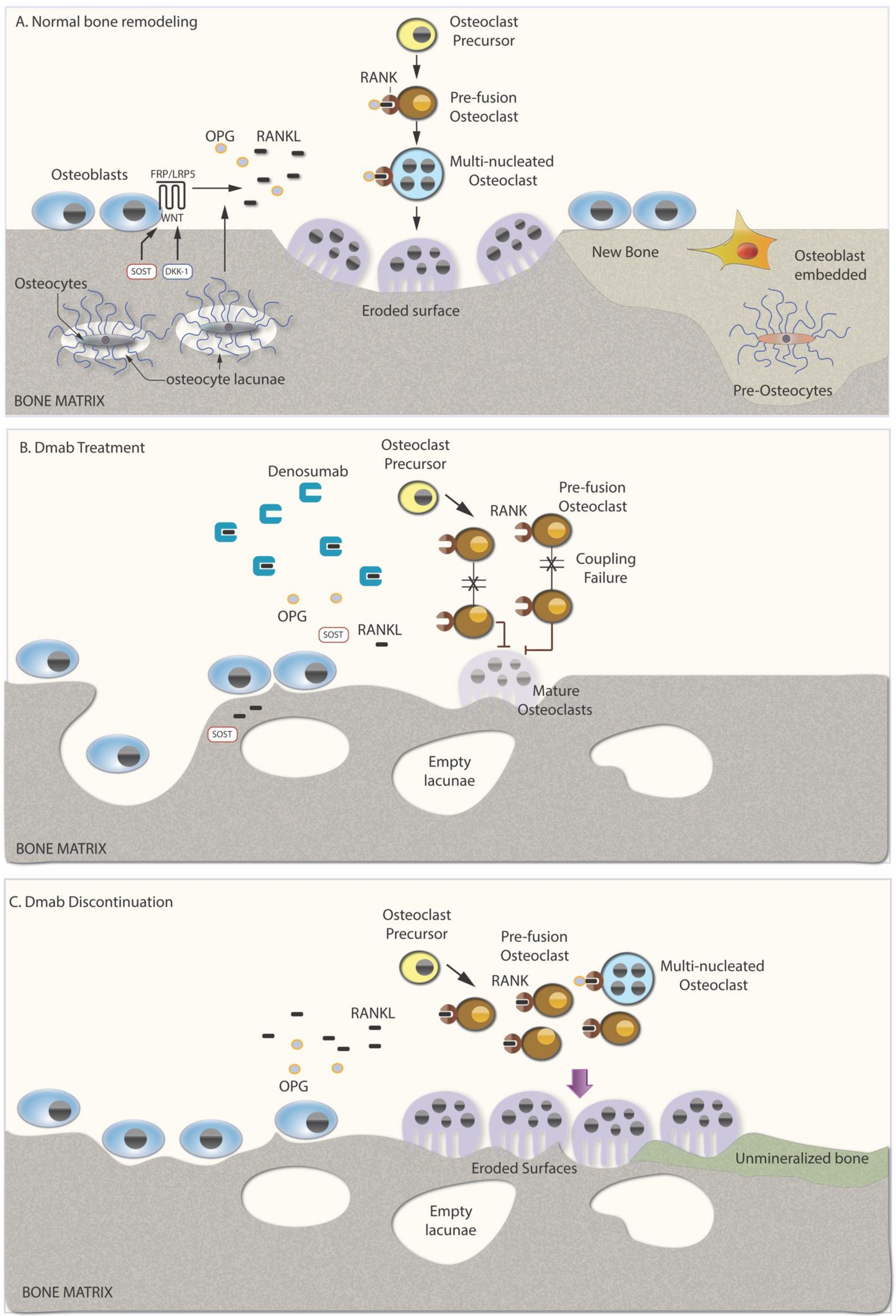
3. Mechanism of Dmab Action—Effect of Discontinuation on Bone Metabolism
4. Discontinuation Effect on Bone Turnover Markers
5. Discontinuation Effect on Bone Mineral Density
6. Discontinuation Effect on Fracture Risk
Main Limitations of the Studies
7. Factors Predisposing to Bone Loss and Fractures Following Discontinuation
8. Patient Management Following Discontinuation
9. Conclusions
10. Recommendations to Clinicians
- Preventing bone loss upon Dmab discontinuation is an issue of concern.
- There are currently limited data and evidence regarding the optimal management of patients discontinuing Dmab. However, this is a matter of ongoing clinical research and new data are continuously emerging.
- BTM should be measured at 3 months after initiation of an oral BP to monitor adherence and efficacy; the maintenance of BTM below the mean of healthy premenopausal women could be considered as an adequate response [61]. In case of zoledronate infusion, BTM measurement should be performed at 3 and 6 months and if values are increased a repeat zoledronate infusion should be considered. Preferable BTM are serum CTX (its concentration should be maintained below 280 ng/L) and PINP (its concentration should be maintained below 35 μg/L) [61].
- BMD testing should be performed at Dmab discontinuation and at 12 months of subsequent antiresorptive treatment [61,83]. A BMD reduction greater than the least significant change (LSC) should be considered an inadequate response both in case of oral and i.v. BPs and would signify the need of either continuation of oral treatment or of an additional zoledronate infusion. Subsequent BMD monitoring should be individualized, depending on each patient’s clinical condition and therapeutic approach [83].
- Spine X-rays and/or vertebral fracture assessment (VFA) should accompany each DXA measurement to identify new VFs.
- According to current evidence, zoledronate infusion or oral alendronate could be preferred as subsequent treatment in patients discontinuing Dmab. No robust data are available for the use of teriparatide.
- The duration of subsequent BP treatment is proposed to last 1–2 years, although this has not been proven in prospective studies.
- The duration of subsequent BP treatment to prevent bone loss may be affected by the duration of Dmab treatment. Physicians should have this in mind when they plan their treatment strategy.
Author Contributions
Funding
Institutional Review Board Statement
Informed Consent Statement
Data Availability Statement
Conflicts of Interest
References
- Anastasilakis, A.D.; Polyzos, S.A.; Makras, P. Therapy of Endocrine Disease: Denosumab vs bisphosphonates for the treatment of postmenopausal osteoporosis. Eur. J. Endocrinol. 2018, 179, R31–R45. [Google Scholar] [CrossRef] [PubMed]
- Anastasilakis, A.D.; Polyzos, S.A.; Yavropoulou, M.P.; Makras, P. Combination and sequential treatment in women with postmenopausal osteoporosis. Expert Opin. Pharmacother. 2020, 21, 477–490. [Google Scholar] [CrossRef] [PubMed]
- Bone, H.G.; Wagman, R.B.; Brandi, M.L.; Brown, J.P.; Chapurlat, R.; Cummings, S.R.; Czerwiński, E.; Fahrleitner-Pammer, A.; Kendler, D.L.; Lippuner, K.; et al. 10 years of denosumab treatment in postmenopausal women with osteoporosis: Results from the phase 3 randomised FREEDOM trial and open-label extension. Lancet Diabetes Endocrinol. 2017, 5, 513–523. [Google Scholar] [CrossRef]
- Bone, H.G.; Bolognese, M.A.; Yuen, C.K.; Kendler, D.L.; Miller, P.D.; Yang, Y.-C.; Grazette, L.; Martin, J.S.; Gallagher, J.C. Effects of Denosumab Treatment and Discontinuation on Bone Mineral Density and Bone Turnover Markers in Postmenopausal Women with Low Bone Mass. J. Clin. Endocrinol. Metab. 2011, 96, 972–980. [Google Scholar] [CrossRef] [PubMed] [Green Version]
- Manolagas, S.C. The Quest for Osteoporosis Mechanisms and Rational Therapies: How Far We’ve Come, How Much Further We Need to Go. J. Bone Miner. Res. 2018, 33, 371–385. [Google Scholar] [CrossRef] [Green Version]
- Manolagas, S.C.; Parfitt, A.M. For whom the bell tolls: Distress signals from long-lived osteocytes and the pathogenesis of metabolic bone diseases. Bone 2013, 54, 272–278. [Google Scholar] [CrossRef] [Green Version]
- Goldring, S.R. The osteocyte: Key player in regulating bone turnover. RMD Open 2015, 1, e000049. [Google Scholar] [CrossRef]
- Tu, X.; Delgado-Calle, J.; Condon, K.W.; Maycas, M.; Zhang, H.; Carlesso, N.; Taketo, M.M.; Burr, D.B.; Plotkin, L.I.; Bellido, T. Osteocytes mediate the anabolic actions of canonical Wnt/β-catenin signaling in bone. Proc. Natl. Acad. Sci. USA 2015, 112, E478–E486. [Google Scholar] [CrossRef] [Green Version]
- Yavropoulou, M.P.; Yovos, J.G. The role of the Wnt signaling pathway in osteoblast commitment and differentiation. Hormones Athens 2007, 6, 279–294. [Google Scholar] [CrossRef]
- Xiong, J.; Piemontese, M.; Onal, M.; Campbell, J.; Goellner, J.J.; Dusevich, V.; Bonewald, L.; Manolagas, S.C.; O’Brien, C.A. Osteocytes, not Osteoblasts or Lining Cells, are the Main Source of the RANKL Required for Osteoclast Formation in Remodeling Bone. PLoS ONE 2015, 10, e0138189. [Google Scholar] [CrossRef]
- Nakashima, T.; Hayashi, M.; Fukunaga, T.; Kurata, K.; Oh-Hora, M.; Feng, J.Q.; Bonewald, L.F.; Kodama, T.; Wutz, A.; Wagner, E.F.; et al. Evidence for osteocyte regulation of bone homeostasis through RANKL expression. Nat. Med. 2011, 17, 1231–1234. [Google Scholar] [CrossRef] [PubMed]
- Solberg, L.B.; Stang, E.; Brorson, S.-H.; Andersson, G.; Reinholt, F.P. Tartrate-resistant acid phosphatase (TRAP) co-localizes with receptor activator of NF-KB ligand (RANKL) and osteoprotegerin (OPG) in lysosomal-associated membrane protein 1 (LAMP1)-positive vesicles in rat osteoblasts and osteocytes. Histochem. Cell Biol. 2015, 143, 195–207. [Google Scholar] [CrossRef] [PubMed] [Green Version]
- Weitzmann, M.N.; Pacifici, R. Estrogen deficiency and bone loss: An inflammatory tale. J. Clin. Investig. 2006, 116, 1186–1194. [Google Scholar] [CrossRef] [PubMed] [Green Version]
- Geusens, P.P.; Landewé, R.B.M.; Garnero, P.; Chen, D.; Dunstan, C.R.; Lems, W.F.; Stinissen, P.; van der Heijde, D.M.F.M.; van der Linden, S.; Boers, M. The ratio of circulating osteoprotegerin to RANKL in early rheumatoid arthritis predicts later joint destruction. Arthritis Rheum. 2006, 54, 1772–1777. [Google Scholar] [CrossRef]
- Tanaka, S.; Tanaka, Y. RANKL as a therapeutic target of rheumatoid arthritis. J. Bone Miner. Metab. 2020. [Google Scholar] [CrossRef]
- Humphrey, E.L.; Williams, J.H.H.; Davie, M.W.J.; Marshall, M.J. Effects of dissociated glucocorticoids on OPG and RANKL in osteoblastic cells. Bone 2006, 38, 652–661. [Google Scholar] [CrossRef]
- Brandi, M.L.; Cavalli, L. Targeted approaches in the treatment of osteoporosis: Differential mechanism of action of denosumab and clinical utility. Ther. Clin. Risk Manag. 2012, 8, 253–266. [Google Scholar] [CrossRef] [Green Version]
- Hanley, D.A.; Adachi, J.D.; Bell, A.; Brown, V. Denosumab: Mechanism of action and clinical outcomes. Int. J. Clin. Pr. 2012, 66, 1139–1146. [Google Scholar] [CrossRef] [Green Version]
- Jähn-Rickert, K.; Wölfel, E.M.; Jobke, B.; Riedel, C.; Hellmich, M.; Werner, M.; McDonald, M.M.; Busse, B. Elevated Bone Hardness Under Denosumab Treatment, With Persisting Lower Osteocyte Viability During Discontinuation. Front. Endocrinol. 2020, 11, 250. [Google Scholar] [CrossRef]
- McClung, M.R. Cancel the denosumab holiday. Osteoporos. Int. 2016, 27, 1677–1682. [Google Scholar] [CrossRef] [Green Version]
- Tsourdi, E.; Zillikens, M.C. Certainties and Uncertainties About Denosumab Discontinuation. Calcif. Tissue Int. 2018, 103, 1–4. [Google Scholar] [CrossRef] [PubMed]
- Brown, J.P.; Dempster, D.W.; Ding, B.; Dent-Acosta, R.; Martin, J.S.; Grauer, A.; Wagman, R.B.; Zanchetta, J. Bone remodeling in postmenopausal women who discontinued denosumab treatment: Off-treatment biopsy study. J. Bone Miner. Res. 2011, 26, 2737–2744. [Google Scholar] [CrossRef] [PubMed]
- Zanchetta, M.B.; Boailchuk, J.; Massari, F.; Silveira, F.; Bogado, C.; Zanchetta, J.R. Significant bone loss after stopping long-term denosumab treatment: A post FREEDOM study. Osteoporos. Int. 2018, 29, 41–47. [Google Scholar] [CrossRef]
- Anastasilakis, A.D.; Polyzos, S.A.; Makras, P.; Aubry-Rozier, B.; Kaouri, S.; Lamy, O. Clinical Features of 24 Patients With Rebound-Associated Vertebral Fractures After Denosumab Discontinuation: Systematic Review and Additional Cases. J. Bone Miner. Res. 2017, 32, 1291–1296. [Google Scholar] [CrossRef] [PubMed] [Green Version]
- Saag, K.; McDermott, M.; Adachi, J.; Lems, W.; Lane, N.; Geusens, P.; Butler, P.; Chen, L.; Crittenden, D.B.; Dore, R.; et al. Effect of Discontinuation of Denosumab in Subjects with Rheumatoid Arthritis Treated with Glucocorticoids. Arthritis Rheumatol. 2019, 71 (Suppl. 10), 1876. [Google Scholar]
- Gonzalez-Rodriguez, E.; Aubry-Rozier, B.; Stoll, D.; Zaman, K.; Lamy, O. Sixty spontaneous vertebral fractures after denosumab discontinuation in 15 women with early-stage breast cancer under aromatase inhibitors. Breast Cancer Res. Treat. 2019, 179, 153–159. [Google Scholar] [CrossRef] [PubMed]
- Miller, P.D.; Bolognese, M.A.; Lewiecki, E.M.; McClung, M.R.; Ding, B.; Austin, M.; Liu, Y.; Martin, J.S. Effect of denosumab on bone density and turnover in postmenopausal women with low bone mass after long-term continued, discontinued, and restarting of therapy: A randomized blinded phase 2 clinical trial. Bone 2008, 43, 222–229. [Google Scholar] [CrossRef]
- McClung, M.R.; Wagman, R.B.; Miller, P.D.; Wang, A.; Lewiecki, E.M. Observations following discontinuation of long-term denosumab therapy. Osteoporos. Int. 2017, 28, 1723–1732. [Google Scholar] [CrossRef] [Green Version]
- Fassio, A.; Adami, G.; Benini, C.; Vantaggiato, E.; Saag, K.; Giollo, A.; Lippolis, I.; Viapiana, O.; Idolazzi, L.; Orsolini, G.; et al. Changes in Dkk-1, sclerostin, and RANKL serum levels following discontinuation of long-term denosumab treatment in postmenopausal women. Bone 2019, 123, 191–195. [Google Scholar] [CrossRef]
- Anastasilakis, A.D.; Yavropoulou, M.P.; Makras, P.; Sakellariou, G.T.; Papadopoulou, F.; Gerou, S.; Papapoulos, S.E. Increased osteoclastogenesis in patients with vertebral fractures following discontinuation of denosumab treatment. Eur. J. Endocrinol. 2017, 176, 677–683. [Google Scholar] [CrossRef]
- Uebelhart, B.; Rizzoli, R.; Ferrari, S.L. Retrospective evaluation of serum CTX levels after denosumab discontinuation in patients with or without prior exposure to bisphosphonates. Osteoporos. Int. 2017, 28, 2701–2705. [Google Scholar] [CrossRef] [PubMed]
- Kobel, C.; Frey, D.; Graf, N.; Wüthrich, R.P.; Bonani, M. Follow-Up of Bone Mineral Density Changes in de novo Kidney Transplant Recipients Treated with Two Doses of the Receptor Activator of Nuclear Factor κB Ligand Inhibitor Denosumab. Kidney Blood Press. Res. 2019, 44, 1285–1293. [Google Scholar] [CrossRef] [PubMed]
- Popp, A.W.; Varathan, N.; Buffat, H.; Senn, C.; Perrelet, R.; Lippuner, K. Bone Mineral Density Changes After 1 Year of Denosumab Discontinuation in Postmenopausal Women with Long-Term Denosumab Treatment for Osteoporosis. Calcif. Tissue Int. 2018, 103, 50–54. [Google Scholar] [CrossRef] [PubMed]
- Cummings, S.; Ferrari, S.; Eastell, R.; Gilchrist, N.; Jensen, J.-E.B.; McClung, M.; Roux, C.; Törring, O.; Valter, I.; Wang, A.T.; et al. Vertebral Fractures After Discontinuation of Denosumab: A Post Hoc Analysis of the Randomized Placebo-Controlled FREEDOM Trial and Its Extension. J. Bone Miner. Res. 2018, 33, 190–198. [Google Scholar] [CrossRef] [PubMed] [Green Version]
- Japelj, M.; Vidmar, G.; Rajic, A.S.; Pfeifer, M.; Kocjan, T. Bone mineral density decline following denosumab discontinuation might not be attenuated with previous bisphosphonate therapy. Endocr. Abstr. 2018, 56, 188. [Google Scholar] [CrossRef]
- Aubry-Rozier, B.; Liebich, G.; Stoll, D.; Gonzalez-Rodriguez, E.; Hans, D.; Lamy, O. Can We Avoid the Loss of Bone Mineral Density One Year After Denosumab Discontinuation? The Reolaus Bone Project. Presented at the Annual European Congress of Rheumatology, EULAR 2019, Madrid, Spain, 12–15 June 2019. [Google Scholar]
- Kendler, D.; Roux, C.; Benhamou, C.L.; Brown, J.P.; Lillestol, M.; Siddhanti, S.; Man, H.-S.; Martin, J.S.; Bone, H.G. Effects of denosumab on bone mineral density and bone turnover in postmenopausal women transitioning from alendronate therapy. J. Bone Miner. Res. 2009, 25, 72–81. [Google Scholar] [CrossRef]
- Lyu, H.; Yoshida, K.; Zhao, S.S.; Wei, J.; Zeng, C.; Tedeschi, S.K.; Leder, B.Z.; Lei, G.; Tang, P.; Solomon, D.H. Delayed Denosumab Injections and Fracture Risk Among Patients With Osteoporosis: A Population-Based Cohort Study. Ann. Intern. Med. 2020, 173, 516–526. [Google Scholar] [CrossRef]
- Tripto-Shkolnik, L.; Fund, N.; Rouach, V.; Chodick, G.; Shalev, V.; Goldshtein, I. Fracture incidence after denosumab discontinuation: Real-world data from a large healthcare provider. Bone 2019, 130, 115150. [Google Scholar] [CrossRef]
- Brown, J.P.; Roux, C.; Törring, O.; Ho, P.R.; Beck Jensen, J.E.; Gilchrist, N.; Recknor, C.; Austin, M.; Wang, A.; Grauer, A.; et al. Discontinuation of denosumab and associated fracture incidence: Analysis from the Fracture Reduction Evaluation of Denosumab in Osteoporosis Every 6 Months (FREEDOM) Trial. J. Bone Miner. Res. 2012, 28, 746–752. [Google Scholar] [CrossRef] [Green Version]
- Anastasilakis, A.D.; Evangelatos, G.; Makras, P.; Iliopoulos, A. Rebound-associated vertebral fractures may occur in sequential time points following denosumab discontinuation: Need for prompt treatment re-initiation. Bone Rep. 2020, 12, 100267. [Google Scholar] [CrossRef]
- Kashii, M.; Ebina, K.; Kitaguchi, K.; Yoshikawa, H. Romosozumab was not effective in preventing multiple spontaneous clinical vertebral fractures after denosumab discontinuation: A case report. Bone Rep. 2020, 13, 100288. [Google Scholar] [CrossRef] [PubMed]
- Anastasilakis, A.D.; Evangelatos, G.; Makras, P.; Iliopoulos, A. Magnetic resonance imaging has an advantage over conventional spine X-rays in the evaluation of rebound-associated vertebral fractures following denosumab discontinuation. Endocrine 2020, 69, 516–518. [Google Scholar] [CrossRef]
- Fernández-Fernández, E.; Benavent-Núñez, D.; Bonilla Hernán, G.; Monjo-Henry, I.; Garcia Carazo, S.; Bernad Pineda, M.; Balsa Criado, A.; Acín, P.A. Multiple vertebral fractures following discontinuation of denosumab treatment: Ten clinical cases report. Reumatl. Clin. 2020, 16, 480–484. [Google Scholar] [CrossRef]
- Florez, H.; Ramírez, J.; Monegal, A.; Guañabens, N.; Peris, P. Spontaneous vertebral fractures after denosumab discontinuation: A case collection and review of the literature. Semin. Arthritis Rheum. 2019, 49, 197–203. [Google Scholar] [CrossRef] [PubMed]
- Anastasilakis, A.D.; Papapoulos, S.E.; Polyzos, S.A.; Appelman-Dijkstra, N.M.; Makras, P. Zoledronate for the Prevention of Bone Loss in Women Discontinuing Denosumab Treatment. A Prospective 2-Year Clinical Trial. J. Bone Miner. Res. 2019, 34, 2220–2228. [Google Scholar] [CrossRef]
- Lamy, O.; Fernández-Fernández, E.; Monjo-Henry, I.; Stoll, D.; Aubry-Rozier, B.; Benavent-Núñez, D.; Aguado, P.; Gonzalez-Rodriguez, E. Alendronate after denosumab discontinuation in women previously exposed to bisphosphonates was not effective in preventing the risk of spontaneous multiple vertebral fractures: Two case reports. Osteoporos. Int. 2019, 30, 1111–1115. [Google Scholar] [CrossRef]
- Anagnostis, P.; Paschou, S.A.; Gonzalez-Rodriguez, E.; Potoupnis, M.; Tsiridis, E.; Lamy, O.; Goulis, D.G. Spontaneous Vertebral Fractures in Males with Osteoporosis After Denosumab Discontinuation: A Report of Two Cases. J. Clin. Rheumatol. 2019. [Google Scholar] [CrossRef]
- De Sousa, S.M.C.; Jesudason, D. Rebound vertebral and non-vertebral fractures during denosumab interruption in a postmenopausal woman. Clin. Endocrinol. 2019, 90, 250–252. [Google Scholar] [CrossRef] [Green Version]
- Gonzalez-Rodriguez, E.; Stoll, D.; Lamy, O. Raloxifene Has No Efficacy in Reducing the High Bone Turnover and the Risk of Spontaneous Vertebral Fractures after Denosumab Discontinuation. Case Rep. Rheumatol. 2018, 2018, 1–4. [Google Scholar] [CrossRef]
- Che, H.; Breuil, V.; Cortet, B.; Paccou, J.; Thomas, T.; Chapuis, L.; Debiais, F.; Mehsen-Cetre, N.; Javier, R.M.; Peres, S.L.; et al. Vertebral fractures cascade: Potential causes and risk factors. Osteoporos. Int. 2018, 30, 555–563. [Google Scholar] [CrossRef]
- Tripto-Shkolnik, L.; Rouach, V.; Marcus, Y.; Rotman-Pikielny, P.; Benbassat, C.; Vered, I. Vertebral Fractures Following Denosumab Discontinuation in Patients with Prolonged Exposure to Bisphosphonates. Calcif. Tissue Int. 2018, 103, 44–49. [Google Scholar] [CrossRef] [PubMed]
- Niimi, R.; Kono, T.; Nishihara, A.; Hasegawa, M.; Sudo, A. Rebound-associated vertebral fractures after discontinuation of denosumab for the treatment of maxillitis. Osteoporos. Int. 2018, 29, 769–772. [Google Scholar] [CrossRef] [PubMed]
- Polyzos, S.A.; Terpos, E. Clinical vertebral fractures following denosumab discontinuation. Endocrine 2016, 54, 271–272. [Google Scholar] [CrossRef]
- Lamy, O.; Gonzalez-Rodriguez, E.; Stoll, D.; Hans, D.; Aubry-Rozier, B. Severe Rebound-Associated Vertebral Fractures After Denosumab Discontinuation: 9 Clinical Cases Report. J. Clin. Endocrinol. Metab. 2017, 102, 354–358. [Google Scholar] [CrossRef] [PubMed]
- Popp, A.W.; Zysset, P.; Lippuner, K. Rebound-associated vertebral fractures after discontinuation of denosumab—From clinic and biomechanics. Osteoporos. Int. 2016, 27, 1917–1921. [Google Scholar] [CrossRef] [PubMed]
- Aubry-Rozier, B.; Gonzalez-Rodriguez, E.; Stoll, D.; Lamy, O. Severe spontaneous vertebral fractures after denosumab discontinuation: Three case reports. Osteoporos. Int. 2016, 27, 1923–1925. [Google Scholar] [CrossRef] [PubMed]
- Anastasilakis, A.D.; Makras, P. Multiple clinical vertebral fractures following denosumab discontinuation. Osteoporos. Int. 2016, 27, 1929–1930. [Google Scholar] [CrossRef]
- Black, D.M.; Arden, N.K.; Palermo, L.; Pearson, J.; Cummings, S.R. Prevalent Vertebral Deformities Predict Hip Fractures and New Vertebral Deformities but Not Wrist Fractures. J. Bone Miner. Res. 1999, 14, 821–828. [Google Scholar] [CrossRef]
- Lindsay, R.; Silverman, S.L.; Cooper, C.; Hanley, D.A.; Barton, I.; Broy, S.B.; Licata, A.; Benhamou, L.; Geusens, P.; Flowers, K.; et al. Risk of New Vertebral Fracture in the Year Following a Fracture. JAMA 2001, 285, 320–323. [Google Scholar] [CrossRef]
- Tsourdi, E.; Zillikens, M.C.; Meier, C.; Body, J.-J.; Gonzalez-Rodriguez, E.; Anastasilakis, A.D.; Abrahamsen, B.; McCloskey, E.; Hofbauer, L.C.; Guañabens, N.; et al. Fracture Risk and Management of Discontinuation of Denosumab Therapy: A Systematic Review and Position Statement by ECTS. J. Clin. Endocrinol. Metab. 2021, 106, 264–281. [Google Scholar] [CrossRef]
- Koldkjær Sølling, A.S.; Harsløf, T.; Kaal, A.; Rejnmark, L.; Langdahl, B. Hypercalcemia after discontinuation of long-term denosumab treatment. Osteoporos. Int. 2016, 27, 2383–2386. [Google Scholar] [CrossRef] [PubMed]
- Sølling, A.S.; Harsløf, T.; Langdahl, B. Treatment with Zoledronate Subsequent to Denosumab in Osteoporosis: A Randomized Trial. J. Bone Miner. Res. 2020, 35, 1858–1870. [Google Scholar] [CrossRef] [PubMed]
- Ferrari, S.; Libanati, C.; Lin, C.J.F.; Brown, J.P.; Cosman, F.; Czerwiński, E.; de Gregόrio, L.H.; Malouf-Sierra, J.; Reginster, J.-Y.; Wang, A.; et al. Relationship Between Bone Mineral Density T-Score and Nonvertebral Fracture Risk Over 10 Years of Denosumab Treatment. J. Bone Miner. Res. 2019, 34, 1033–1040. [Google Scholar] [CrossRef] [Green Version]
- Cummings, S.; Cosman, F.; Lewiecki, E.M.; Schousboe, J.T.; Bauer, D.C.; Black, D.M.; Brown, T.D.; Cheung, A.M.; Cody, K.; Cooper, C.; et al. Goal-Directed Treatment for Osteoporosis: A Progress Report From the ASBMR-NOF Working Group on Goal-Directed Treatment for Osteoporosis. J. Bone Miner. Res. 2017, 32, 3–10. [Google Scholar] [CrossRef] [PubMed]
- Anastasilakis, A.D.; Trovas, G.; Balanika, A.; Polyzos, S.A.; Makras, P.; Tournis, S. Progression of Rebound-Associated Vertebral Fractures Following Denosumab Discontinuation Despite Reinstitution of Treatment: Suppressing Increased Bone Turnover May Not Be Enough. J. Clin. Densitom. 2020, in press. [Google Scholar] [CrossRef] [PubMed]
- Leder, B.Z.; Tsai, J.N.; Uihlein, A.V.; Wallace, P.M.; Lee, H.; Neer, R.M.; Burnett-Bowie, S.A.M. Denosumab and teriparatide transitions in postmenopausal osteoporosis (the DATA-Switch study): Extension of a randomised controlled trial. Lancet 2015, 386, 1147–1155. [Google Scholar] [CrossRef] [Green Version]
- Cosman, F.; McMahon, D.; Dempster, D.; Nieves, J. Standard Versus Cyclic Teriparatide and Denosumab Treatment for Osteoporosis: A Randomized Trial. J. Bone Miner. Res. 2020, 35, 219–225. [Google Scholar] [CrossRef]
- Ebina, K.; Miyama, A.; Hirao, M.; Yoshikawa, H.; Hashimoto, J.; Kashii, M.; Nakaya, H.; Takahi, K.; Tsuji, S.; Tsuboi, H. Assessment of the effects of sequential treatment after discontinuing denosumab in 64 patients with postmenopausal osteoporosis. J. Bone Miner. Res. 2019, 34, S259. [Google Scholar]
- Freemantle, N.; Satram-Hoang, S.; Tang, E.T.; Kaur, P.; Macarios, D.; Siddhanti, S.; Borenstein, J.; Kendler, D.L. Final results of the DAPS (Denosumab Adherence Preference Satisfaction) study: A 24-month, randomized, crossover comparison with alendronate in postmenopausal women. Osteoporos. Int. 2011, 23, 317–326. [Google Scholar] [CrossRef] [Green Version]
- Kendler, D.; Chines, A.; Clark, P.; Ebeling, P.R.; McClung, M.; Rhee, Y.; Huang, S.; Stad, R.K. Bone Mineral Density After Transitioning From Denosumab to Alendronate. J. Clin. Endocrinol. Metab. 2020, 105. [Google Scholar] [CrossRef] [Green Version]
- Horne, A.M.; Mihov, B.; Reid, I.R. Bone Loss After Romosozumab/Denosumab: Effects of Bisphosphonates. Calcif. Tissue Int. 2018, 103. [Google Scholar] [CrossRef] [PubMed]
- Laroche, M.; Couture, G.; Ruyssen-Witrand, A.; Constantin, A.; Degboé, Y. Effect of risedronate on bone loss at discontinuation of denosumab. Bone Rep. 2020, 13, 100290. [Google Scholar] [CrossRef] [PubMed]
- Zanchetta, M.; Pelegrin, C.; Silveira, F.; Bogado, C.; Zanchetta, J.; Salerni, H.; Costanzon, P. Bisphosphonates prevent bone loss associated with denosumab discontinuation. J. Bone Miner. Res. 2019, 34, 114–115. [Google Scholar]
- Lehmann, T.; Aeberli, D. Possible protective effect of switching from denosumab to zoledronic acid on vertebral fractures. Osteoporos. Int. 2017, 28, 3067–3068. [Google Scholar] [CrossRef] [PubMed]
- Leder, B.Z.; Tsai, J.N.; Jiang, L.A.; Lee, H. Importance of prompt antiresorptive therapy in postmenopausal women discontinuing teriparatide or denosumab: The Denosumab and Teriparatide Follow-up study (DATA-Follow-up). Bone 2017, 98, 54–58. [Google Scholar] [CrossRef]
- Reid, I.R.; Horne, A.M.; Mihov, B.; Gamble, G.D. Bone Loss After Denosumab: Only Partial Protection with Zoledronate. Calcif. Tissue Int. 2017, 101, 371–374. [Google Scholar] [CrossRef]
- Everts-Graber, J.; Reichenbach, S.; Ziswiler, H.R.; Studer, U.; Lehmann, T. A Single Infusion of Zoledronate in Postmenopausal Women Following Denosumab Discontinuation Results in Partial Conservation of Bone Mass Gains. J. Bone Miner. Res. 2020, 35, 1207–1215. [Google Scholar] [CrossRef]
- Everts-Graber, J.; Reichenbach, S.; Gahl, B.; Ziswiler, H.; Studer, U.; Lehmann, T. Risk factors for vertebral fractures and bone loss after denosumab discontinuation: A real-world observational study. Bone 2021, 144, 115830. [Google Scholar] [CrossRef]
- Kondo, H.; Okimoto, N.; Yoshioka, T.; Akahoshi, S.; Fuse, Y.; Ogawa, T.; Okazaki, Y.; Katae, Y.; Tsukamoto, M.; Yamanaka, Y.; et al. Zoledronic acid sequential therapy could avoid disadvantages due to the discontinuation of less than 3-year denosumab treatment. J. Bone Miner. Metab. 2020, 38, 894–902. [Google Scholar] [CrossRef]
- Makras, P.; Papapoulos, S.E.; Polyzos, S.A.; Appelman-Dijkstra, N.M.; Anastasilakis, A.D. The three-year effect of a single zoledronate infusion on bone mineral density and bone turnover markers following denosumab discontinuation in women with postmenopausal osteoporosis. Bone 2020, 138, 115478. [Google Scholar] [CrossRef]
- Anastasilakis, A.D.; Polyzos, S.A.; Yavropoulou, M.P.; Appelman-Dijkstra, N.M.; Ntenti, C.; Mandanas, S.; Papatheodorou, A.; Makras, P. Comparative effect of zoledronate at 6 versus 18 months following denosumab discontinuation. Calcif Tissue Int. 2021. [Google Scholar] [CrossRef] [PubMed]
- Kendler, D.L.; Compston, J.; Carey, J.J.; Wu, C.-H.; Ibrahim, A.; Lewiecki, E.M. Repeating Measurement of Bone Mineral Density when Monitoring with Dual-energy X-ray Absorptiometry: 2019 ISCD Official Position. J. Clin. Densitom. 2019, 22, 489–500. [Google Scholar] [CrossRef] [PubMed]


| Author, Journal, Ref. | Year | Type of Study | n. of Patients | Population | Previous Bone Active Treatment | Time from Last Injection to VF | Treatment Duration with Dmab | Number of Fractures/Risk of Fractures | VF before Treatment | Other Bone Active Treatment After | Cause of Discontinuation |
|---|---|---|---|---|---|---|---|---|---|---|---|
| Houchen Lyu, Annals of Internal Medicine [38] | 2020 | Observational | 2594 | Mean age: 76 y (SD, 10). 94% female, 53% had a history of major osteoporotic fracture. | NA | delay by more than 16 weeks | NA | HR 3.91 (CI, 1.62 to 9.45). | 15% of population | NA | NA |
| Anastasilakis, Bone Rep. [41] | 2020 | case series | 3 | Case 1: 71 y woman naïve to treatment; Case 2: 76 y woman treated with ibandronate for 5 y and treated with GCs; Case 3: 53 y male treated with GCs and alendronate for 3 y. | see population | Case 1: 8 months and 10 months; Case 2: 8 months and 17 months; Case 3: 3 months and 2 months of delay in the injection | Case 1: 8 y; Case 2: 2 y; Case 3: 3 y | Case 1: 2; Case 2: 5; Case 3: 5 | no | Case 1: no; Case 2: no; Case 3: Dmab | Case 1: dentist advice; Case 2: NA; Case 3: patient discontinued |
| Kashii, Bone reports [42] | 2020 | case report | 1 | 60 y woman with osteoporosis complicated by 2 VFs (T6 and T8). | none | 12 months | 5 doses | 5 (T12, L2, L3, L4, and L5) | 2 (T6 and T8) | romosozumab 210 monthly, 9 months after last Dmab (fractures occurred after 3 doses of romosozumab | patient negligence |
| Anastasilakis, Endocrine [43] | 2020 | Case reports | 2 | Case 1: 66 y woman previously treated with alendronate for 1 y; Case 2: 52 y postmenopausal woman with rheumatoid arthritis treated with GCs and methotrexate. | Case 1: alendronate; Case 2: no | Case 1: 9 months; Case 2: 9 months | Case 1: 10 doses; Case 2: 9 doses | Case 1: T10, T11, L3, L4; Case 2: L1 | Case 1: no; Case 2: no | Case 1: na; Case 2: Dmab | Case 1: physician decision (became osteopenic); Case 2: patient negligence |
| Tripto-Shkolnik, Bone [39] | 2020 | retrospective | 1500 | Subjects at least 1 y of treatment. 92% females, mean age = 71.8 ± 9.5 y. Only clinical fractures. | NA | 3 months or more | >2 doses | Multiple VFs occurred in 12 (0.8%). The overall rate of fractures per 100 patient-years of follow-up was RR 3.2, (2.2–4.89) the rate of VF RR 4.7, (2.3–9.6) and multiple VF RR 14.6, (3.3–65.3, effect size 1.06). | 17% | NA | NA |
| Gonzalez-Rodriguez E.Breast Cancer Research and Treatment [26] | 2019 | case series | 15 | 15 women with early-stage breast cancer treated with AI and denosumab 62.3 ± 7.0 years. | NA | 7 to 16 months after last denosumab injection (mean 10.9 ± 2.0) | 8.2 ± 2.0 doses | from 1 to 11 | only 1 patient | Dmab or BPS | 10: end of the AI treatment; 1 osteopenia; 2 delayed for dental treatments; 1 omitted; and 1 stopped by the patient. |
| Fernández Fernández, Reumatol Clin. [44] | 2020 | restrospective | 10 | 10 women with postmenopausal osteoporosis (66 ± 7.7 y). | 90% of population (7 oral BPS, 5 strontium ranelate, 2 raloxifene, 1 tibolone and 1 calcitonine) | 8–18 months (10.9 ± 3.3months) | 3 to 9 doses, (mean 6 ± 1.7) | 2–9 | 4 patients | TPD: 30%, BPs: 20% Dmab: 20% | 2 dental work, 1 low risk of fracture, 7 termination of the time set by the prescribing doctor. |
| Florez H, Seminars in Arthritis and Rheumatism [45] | 2019 | case series | 7 | 7 patients with postmenopausal osteoporosis (2 GC) median age was 64 y (56–75 y); 4 patients had previous fragility fractures (2 VF). | 5 patients:1 zoledronate1 HRT + BPS2 BPS1 HRT | 10 months (8–20) | 24–53 months (median 38) | 5 (2–8) | 2 | 3 Dmab, 1 combined Dmab and TPD, 3 BPS | 2 dental indication; 1 BMD improvement; 1 poor adherence;3 treatment omission and/or delay. |
| Anastasilakis, JBMR [46] | 2019 | RCT | 57 | Treatment naïve postmenopausal women treated with Dmab and achieved osteopenia were randomized (1:1) to receive a single infusion of ZOL (n = 27, given 6 months after the last Dmab injection) or to continue Dmab (n = 30) for 1 year. Follow up until 2 y from randomization. | none | 18 months (Zol group), 9 months (Dmab group), 12 months (Dmab group) | 2.0 ± 0.2 in Dmab group 2.4 ± 0.2 in ZOL group | 2 subjects: 1 new VF and 1 worsening of previous VFs; 1 subject 1 new VF and 2 worsening of previous VF | yes | NA | Osteopenia (design of the trial) |
| Lamy, osteoporosis International [47] | 2019 | Case reports | 2 | Case 1: 67 y woman treated with risedronate then raloxifene, then Dmab, then alendronate, finally ZOL; Case 2: 68 y woman treated with BPS for 3 y then strontium ranelate finally Dmab for 3 y then alendronate. | Case 1: risedronate for 4 years, then raloxifene for 6 years; Case 2: BPS for 3 years and strontium ranelate for 2.5 years | Case 1: Between 7 and 11 months; Case 2: 8 months and 15 months | Case 1: 7 doses; Case 2: 6 doses | Case 1: T8, T9, and L1; Case 2: T5, T6, T8, T9, T11, L3, then also T7 | Case 1: no; Case 2: 5 (T12, L1, L2, T10 and L4) | Case 1: alendronate and ZOL; Case 2: alendronate then TPD | Case 1: BMD gain and treatment duration; Case 2: osteopenic |
| Anagnostis P, Journal of Clinical Rheumatology [48] | 2019 | letter to the editor | 2 | Case 1: 45 y male with osteoporosis complicated by multiple vertebral low-energy fractures (T7;T10-L5); Case 2: 80y man with osteoporosis complicated by VFs (T9 and L1 to L5) in the context of GCs treatment for polymyalgia rheumatica. | Case 1: TPD; Case 2: none | Case 1: 12 months; Case 2: 14 months | Case 1: 3 doses; Case 2: 2.6 doses | Case 1: 3; Case 2: 2 | Case 1: 9; Case 2: 6 | Case 1: Dmab; Case 2: ibadronate then Dmab again | Case 1: patient omission for musculoskeletal pain; Case 2: general practitioner switch to ibandronate because osteopenia |
| De Sousa SMC, Clin Endocrinology [49] | 2019 | case report | 1 | 70 y woman with postmenopausal osteoporosis and no prior fractures. | Combined hormone replacement for eight years; risedronate for eight years, including one year of concomitant treatment with raloxifene; strontium for two years. | 7 and 8 months | 7 doses | 1 after 7 month, 1 after 8 months, 2 after 10.5 months and also rib fractures | 0 | Dmab after 9 months (3 months delay) | dental issue |
| Gonzalez-Rodriguez, Case Reports in Rheumatology [50] | 2018 | Case report | 1 | 60 y woman in AI therapy with letrozole. | none | 10 months | 12 doses | T11 and L5 | no | raloxifene then Dmab again after VFs | end of AI and osteopenia |
| Che H., Osteoporosis International [51] | 2018 | Restrospective | 8 | Subject with VF cascade defined as 3 or more VF in 1 year. 8 patients after Dmab, in a pool of 135 patients. | 7 BPS | 5 subjects: 4–6 months and 3 subjects: 14–18 months | NA | multiple VF | 5 patients | NA | NA |
| Tripto-Shkolnik, Calcified Tissue International [52] | 2018 | phone survey | 5 | 9 female 74.2 ± 5.3 years. | 6 BPS, 1 tpd plus BPS | 6.5 ± 4.7 months | 4.9 ± 1.6 doses | 36 in 9 patients | 6 patients | Dmab or zol | 4: physician decision; 1: administrative; 3 non-osteoporosis related medical condition; 1 unknown |
| R. Niimi, Osteoporosis International [53] | 2017 | case report | 1 | 69 y woman with severe osteoporosis (L1 fracture) | naïve | 10 months | 5 doses | 5 | 1 | Dmab | Maxillitis |
| Cummings, JBMR [34] | 2017 | RCT | 1471 | Among 1001 participants who discontinued treatment during FREEDOM or Extension with >7 months of follow-up after the last dose (1001, denosumab; 470, placebo). | At least 1 year washout from eventually previous treatment. | >7 months | 2–19 doses | 56 subjects, 36 (61%) multiple VF, 23 subjects non-VF | 24% of population | 14% received BPS | End of study period |
| Anastasilakis, Popp, Polyzos, Lamy, Aubry-Rozier), JBMR [54,55,56,57,58] | 2016–2017 | case series | 24 | 24 postmenopausal women (age 64.1 (48–83)) | strontium ranelate and raloxifene; (n1)TPD(n1), and BPS (n2) | 11.2 (8–16) months | 6 doses (2–10) | 4.7 (1–9) | 33% of population | 5 patients were subjected to vertebroplasty, all unsuccessful. TPD was the most commonly prescribed alone or in combination with Dmab or Dmab alone | 13 osteopenic; 1 end of AI; 5 patient’s wish or negligence; 1 dental issue; 3 Dmab treatment duration |
| Study, Year | Study Type, | n (% Female) | Dmab Duration | Post-Dmab Antiresorptive Regimen | % LS BMD Change (% Mean Dmab Gain Preserved) (% pts Preserved BMD **) | % TH BMD Change (% Mean Dmab Gain Preserved) (% pts Preserved BMD **) | % FN BMD Change (% Mean Dmab Gain Preserved) (% pts Preserved BMD **) | VFs | Non-VFs | Comments |
|---|---|---|---|---|---|---|---|---|---|---|
| Freemantle, 2012 [70] | RCT | 115 (100) | 1 y | ALN, 1 y | 0.6 (100) (NR) | 0.4 (100) (NR) | −0.1 (100) (NR) | 0 | 1 humerus | − DAPS study—primary aim: compliance |
| Lehmann, 2017 [75] | Case series | 22 (100) | 2.5 y | ZOL, 1 infusion | −3.8 (61.2) (NR) | −1.7 (56.4) (NR) | −0.6 (73,9) (NR) | 0 | 1 calcaneous | − BMD measured 2.5 yrs after ZOL |
| Leder, 2017 [76] | Follow-up of RCT | 28 (100) | 2 or 4 y | 1 y, ZOL (n = 8); ALN (n = 8); IBN (n = 2); Dmab (n = 10) | −1.2 (NR) (NR) | NR (NR) (NR) | −0.6 (NR) (NR) | 0 | 1 tibia (stress) | − DATA follow-up − 36% of pts received Dmab |
| Reid, 2017 [77] | Case series | 6 (100) | 7 y | ZOL, 1 infusion | −9.2 (50.3) (NR) | NR (NR) (NR) | NR (NR) (NR) | NR | NR | − Follow-up of FREEDOM pts − BMD reported 2 y after ZOL |
| Horne, 2018 [72] | Case series | 16 (100) | 2 y | 1 y, ZOL (n = 11), RIS (n =5) | ZOL: −5 (73) (NR) RIS: −9.9 (41) (NR) | ZOL: −1.5 (87) (NR) RIS: −3.9 (64) (NR) | NR (NR) (NR) | NR | NR | − Follow-up of FRAME pts (1 y romosozumab or placebo before Dmab) − ZOL was given with up to 6 mo delay |
| Anastasilakis, 2019 [46] | RCT | 27 (100) | 2.4 y | ZOL, 1 infusion | 12 mo: 1.7 (100) (NR) 24 mo: 0.1 (100) (11.1) | NR (NR) (NR) | 12 mo: NR (100) (NR) 24 mo: NR (100) (14.8) | 1 | 0 | − After Dmab study |
| Everts-Graber, 2020 [78] | Retrospective observational | 120 (100) | 2–5 y (mean 3 y) | ZOL, 1 infusion | −3.3 (66) | −2.2 (49) | −1.5 (57) | 3 | 4 (1 calcaneus, low energy—1 distal radius, low energy—1 pubic, high energy—1 humerus, high energy) | − BMD measured 2.5 y after ZOL |
| Everts-Graber, 2020 [79] | Retrospective | 193 (100) | mean 2.5 y | ZOL 1 infusion (n = 171), OR Other (n = 22) (IBN (n = 6), ALN (n = 10), SERMs (n = 6)) | ZOL: −3.6 (NR) (NR) Other: −3.2 (NR) (NR) | ZOL: −2.5 (NR) (NR) Other: −3.4 (NR) (NR) | ZOL: −1.6 (NR) (NR )Other: −3.4 (NR) (NR) | 5 (3 on ZOL, 1 on IBN, 1 on SERM) | 3 (all on ZOL) | − The 120 pts of the previous study were included to this study − BMD measured 1–4 y (median 26 mo) after ZOL |
| Kendler, 2020 [71] | Post-hoc analysis of RCT (see DAPS above) | 115 (100) | 1 y | ALN, 1y | 0.6 (100) (84.1) | 0.4 (100) (92.4) | −0.1 (100) (78.3) | 0 | 1 humerus | |
| Kondo, 2020 [80] | Retrospective observational | 30 (96.7) | <3 y (average 1.5 y) | ZOL, 1 infusion | 1.8 (100) (NR) | NR (NR) (NR) | 2.1 (100) (NR) | 0 | 0 | |
| Laroche, 2020 [73] | 18 (100) | 1–4 y (mean 39 mo) | RIS, 3 mo + 9 mo follow-up without RIS | −4.6 (NR) (NR) | −1.8 (NR) (NR) | NR (NR) (NR) | 1 | 0 | ||
| Makras, 2020 [81] | Extension of RCT (see afterDmab above) | 23 (100) | 2.4 y | ZOL, 1 infusion | 36mo: −1.75 (100) (82.6) | NR (NR) (NR) | 36 mo: NR (100) (95.6) | 0 | 1 metatarsal | − In 4 pts LS BMD and in 1 pt FN BMD decreased to T-score < −2.5 |
| Solling, 2020 [63] | RCT | 59 (88.5) | 4.6 y | ZOL, 1 infusion 6 mo after last Dmab dose (6M) OR 9 mo after last Dmab dose (9M) or when turnover increased (OBS) | 6M: −4.8 (NR) (65) 9M: −4.1 (NR) (65 )OBS: −4.7 (NR) (63) | 6M: −2.6 (NR) (15) 9M: −3.2 (NR) (35) OBS: −3.6 (NR) (37) | 6M: −3.0 (NR) (20) 9M: −3.5 (NR) (30) OBS: −4.6 (NR) (37) | 2 | 2 (1 rib, low energy—1 humerus, high energy fracture) |
Publisher’s Note: MDPI stays neutral with regard to jurisdictional claims in published maps and institutional affiliations. |
© 2021 by the authors. Licensee MDPI, Basel, Switzerland. This article is an open access article distributed under the terms and conditions of the Creative Commons Attribution (CC BY) license (http://creativecommons.org/licenses/by/4.0/).
Share and Cite
Anastasilakis, A.D.; Makras, P.; Yavropoulou, M.P.; Tabacco, G.; Naciu, A.M.; Palermo, A. Denosumab Discontinuation and the Rebound Phenomenon: A Narrative Review. J. Clin. Med. 2021, 10, 152. https://doi.org/10.3390/jcm10010152
Anastasilakis AD, Makras P, Yavropoulou MP, Tabacco G, Naciu AM, Palermo A. Denosumab Discontinuation and the Rebound Phenomenon: A Narrative Review. Journal of Clinical Medicine. 2021; 10(1):152. https://doi.org/10.3390/jcm10010152
Chicago/Turabian StyleAnastasilakis, Athanasios D., Polyzois Makras, Maria P. Yavropoulou, Gaia Tabacco, Anda Mihaela Naciu, and Andrea Palermo. 2021. "Denosumab Discontinuation and the Rebound Phenomenon: A Narrative Review" Journal of Clinical Medicine 10, no. 1: 152. https://doi.org/10.3390/jcm10010152